Bring imaging to life: confocal imaging advanced to levels previously unseen.
Nikon’s groundbreaking confocal imaging system offering greater speed, higher resolution and unprecedented system flexibility.
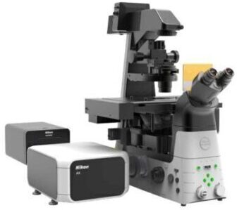
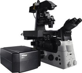
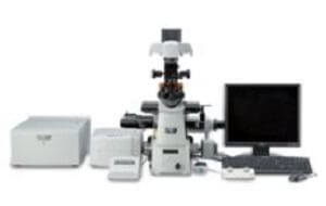
The A1+ confocal laser microscope system is Nikon’s powerful fully-automated confocal imaging system, capable of capturing high-quality confocal images of cells and molecular events at high speed and enhanced sensitivity. Ideal for facilities with a broad range of users, the A1+ has been designed with groundbreaking new optical and electronic technology innovations to provide unprecedented system quality and flexibility. New features such as Nikon’s GaAsP multi-detector unit enables brighter, even higher resolution images than ever before.
ER: Enhance Your Confocal Resolution
The A1 ER enhanced resolution Module combines powerful GPU-based processing and specialized deconvolution algorithms to dramatically enhance the spatial resolution of your A1+ or A1R+ confocal microscope with minimal processing time.
The A1 ER Module provides high quality PSF models for Nikon’s high performance objective lenses, taking the guesswork out of deconvolution analysis. Other features include automatic or manual mode for iteration selection, enhanced spherical aberration correction, and robust algorithms for noise estimation and removal.
Galvano Scanner Enables High-resolution Imaging
The A1+ utilizes a galvano scanner which enables high-resolution imaging of up to 4096 x 4096 pixels. In addition, with the newly developed scanner driving and sampling systems, plus image correction technology, high-speed acquisition of 10 fps (512 x 512 pixels) is also possible.
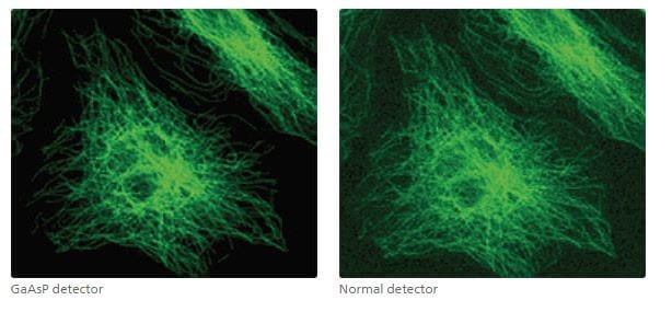
GaAsP Multi Detector Unit
Nikon developed the GaAsP multi-detector unit equipped with two GaAsP PMTs and two normal PMTs.
A GaAsP PMT has much higher sensitivity than a normal PMT, thus acquisition of brighter signals with minimal background noise is possible with a GaAsP PMT, even with weak fluorescence, which, until now, has been difficult to detect.
When using resonant scanners, the GaAsP PMT enables low-noise, high-speed imaging.
Increased Light Detection Efficiency
The low-angle incidence method utilized on the dichroic mirrors increases fluorescence efficiency by 30%.
| Conventional 45° incidence angle method |
Nikon’s original dual integration signal processing technology (DISP) has been implemented in the image processing circuitry to improve electrical efficiency, resulting in an extremely high S/N ratio.
Enhanced Spectral Imaging
Acquisition of a 32-channel spectral image (512 x 512 pixels) with a single scan in 0.6 second is possible. Moreover, 512 x 32-pixel images can be captured at 24 fps.
Accurate, High-speed Unmixing
Accurate spectral unmixing provides maximum performance in the separation of closely overlapping fluorescence spectra and the elimination of autofluorescence. Superior algorithms and high-speed data processing enable real time unmixing during image acquisition.
V-filtering Function
Filter-less intensity adjustment is possible by selecting desired spectral ranges from 32 channels that match the spectrum of the fluorescence probe in use and combining them to perform the filtering function.
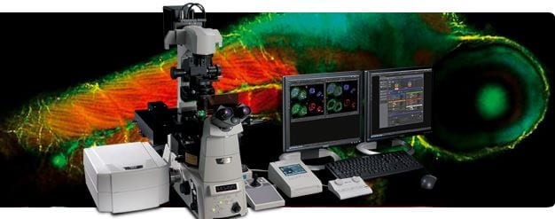
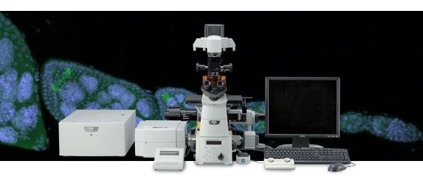
NIS-Elements C control software enables integrated control of the confocal imaging system, microscope and peripheral devices with a simple and intuitive interface. Diverse reliable analysis functions are also available.
| Scan head input/output port | 2 laser input ports 3 signal output ports for standard, spectral and optional detector *1 |
|
|---|---|---|
| Laser | LU-N3 3-laser unit | 405 nm, 488 nm, 561nm lasers are installed ; built-in AOTF *Cannot be used with spectral detector |
| LU-N4/LU-N4S 4-laser unit |
405 nm, 488 nm, 561nm,640nm laser are installed;built-in AOTF *Use LU-N4S when using the spectral detector | |
| LU-NV series laser unit | Compatible lasers : 405 nm, 445 nm, 458nm, 488nm, 514nm, 532nm, 561nm, 594nm, 640nm, 647nm; built-in AOTF | |
| Standard fluorescence detector | Wavelength | 400-750 nm |
| Detector | A1-DU4 4 Detector Unit: 4 standard PMTs A1-DUG GaAsP Multi Detector Unit: 2 GaAsP PMTs + 2 standard PMTs | |
| Filter cube | 6 filter cubes commonly used for a microscope mountable on each of three filter wheels Recommended wavelengths: 450/50, 482/35, 515/30, 525/50, 540/30, 550/49, 585/65, 595/50, 700/75 | |
| Diascopic detector (option) | Wavelength | 485-650 nm |
| Detector | PMT | |
| FOV | Square inscribed in a ø18 mm circle | |
| Image bit depth | 4096 gray intensity levels (12 bit) | |
| Scan head | Standard image acquisition | Scanner: galvano scanner x2 Pixel size: max. 4096 x 4096 pixels Scanning speed: Standard mode: 2 fps (512 x 512 pixels, bi-direction), 24 fps (512 x 32 pixels, bi-direction) Fast mode: 10 fps (512 x 512 pixels, bi-direction), 130 fps (512 x 32 pixels, bi-direction)*2 Zoom: 1-1000x continuously variable Scanning mode: X-Y, X-T, X-Z, XY rotation, Free line |
| High-speed image acquisition | — | |
| Dichroic mirror | Low-angle incidence method, Position: 8 Standard filter: 405/488, 405/488/561, 405/488/561/638, 405/488/543/638, 457/514, BS20/80 Optional filter: 457/514/561 | |
| Pinhole | 12-256 µm variable (1st image plane) | |
| Spectral detector*3 (option) |
Number of channels | 32 channels |
| Wavelength detection range | 400-750 nm | |
| Spectral image acquisition speed | 4 fps (256 x 256 pixels), 1000 lps Pixel size: max. 2048 x 2048 | |
| Wavelength resolution | 80 nm (2.5 nm), 192 nm (6 nm), 320 nm (10 nm) Wavelength range variable in 0.25 nm steps | |
| Unmixing | High-speed unmixing, Precision unmixing | |
| Z step | Ti-E: 0.025 µm, FN1 stepping motor: 0.05 µm Ni-E: 0.025 µm | |
| Compatible microscopes | ECLIPSE Ti-E inverted microscope, ECLIPSE FN1 fixed stage microscope, ECLIPSE Ni-E upright microscope (focusing nosepiece type and focusing stage type) | |
| Option | Motorized XY stage (for Ti-E/Ni-E), High-speed Z stage (for Ti-E), High-speed piezo objective-positioning system (for FN1/Ni-E) | |
| Software | Display/image generation | 2D analysis, 3D volume rendering/orthogonal, 4D analysis, spectral unmixing |
| Image format | JP2, JPG, TIFF, BMP, GIF, PNG, ND2, JFF, JTF, AVI, ICS/IDS | |
| Application | FRAP, FLIP, FRET(option), photoactivation, three-dimensional time-lapse imaging, multipoint time-lapse imaging, colocalization | |
| Control computer | OS | Microsoft Windows® 7 Professional 64bits SP1 |
| CPU | Intel Xeon E5-2643v3 (3.40 GHz/20 MB) or higher | |
| Memory | 16 GB or higher | |
| Hard disk | 300 GB SAS (15,000 rpm) x2, RAID 0 configuration | |
| Data transfer | Dedicated data transfer I/F | |
| Network interface | 10/100/1000 Gigabit Ethernet x2 | |
| Monitor | 1600 x 1200 or higher resolution, dual monitor configuration recommended | |
| Recommended installation conditions | Temperature 23 ± 5 ºC, humidity 70 % (RH) or less (non-condensing) | |
* 1 FCS/FCCS/FLIM is possible in combination with third-party systems
* 2 Fast mode is compatible with zoom 8-1000x and scanning modes X-Y and X-T. It is not compatible with Rotation, Free line, CROP, ROI, Spectral imaging, Stimulation and FLIM.
* 3 Compatible with galvano scanner only.
 Biomechanics
 Biophysics
 Cardiovascular Research
 Dermatology
 Developmental Biology/Embryology
 Endocrinology
 Food Science
 Hepatology
 Molecular Biology
 Nephrology
 Neurobiology / Neuroscience
 Obstetrics & Gynaecology
 Odontology (dentistry)
 Oncology
 Orthopaedics
 Osteology
 Pathology
 Pharmaceutical Sciences
 Rheumatology
 Tropical Medicine
 Urology
 Vascular Research