High-definition macro to micro confocal imaging
With high-definition wide field of view images larger than 1cm with unprecedentedly high S/N ratios, the AZ-C2+ allows for imaging of whole-mount specimens, such as embryos, in a single shot. It offers a combination of low and high magnification objective lenses, optical zoom and a confocal scanning zoom function, enabling continuous imaging from macro to micro. Moreover, the AZ-C2+ allows deep imaging of in vivowhole specimens.
High-definition macro to micro confocal imaging
The AZ-C2+ enables high-definition confocal imaging during macro observation as well as micro observation.
Sharp wide field of view images with unprecedentedly high S/N ratios allow for imaging of whole-mount specimens such as embryos and large tissue slices that are commonly used in developmental biology andsystems biology studies.
Moreover, the AZ-C2+ offers a combination of low and high magnification objective lenses and a scanning zoom function, enabling continuous imaging from macro to micro with a single microscope.
The macro in vivo imaging capabilities allow for the capture of confocal images that were previously not possible with traditional stereoscopic microscopes.
 TT2 ES cells Anti-Nanog antibody (Cy3), anti-Oct3/4 antibody (Alexa488) and DAPI localized in cell nuclei Photographed with the cooperation of: Hiroshi Kiyonari, Laboratory for Animal Resources and Genetic Engineering, RIKEN Center for Developmental Biology |
 Photo courtesy of: Director and Professor Masatoshi Yamamoto, Drosophila Genetic Resource Center, Kyoto Institute of Technology |
Comparison of same specimen regions captured by the AZ-C2+ and an epi-fluorescence microscope
The AZ-C2+ eliminates out-of-focus light and flare to deliver highly resolved confocal fluorescence images and optical sections.
 |
 |
 |
 |
|
Confocal fluorescence
|
Standard wide-field
|
Confocal fluorescence
|
Standard wide-field
|
Specimen: 7.5-day-old mouse embryo expressing H2B-EGFP.
|
Specimen: Zebrafish eye double-stained with GFP and mCherryPhotos courtesy of: 2008 Physiology Course, Marine Biological Laboratory |
||
One-shot whole specimen macro confocal imaging
High NA objectives for macro observation enable fast, high-resolution, single-image capture of a wide specimen area. Because the objectives cover a field of view larger than 1 cm, imaging of embryos during late stages of development and the dynamics of cell populations in whole organs are possible.
[Note] When Plan Apochromat 1x and AZ100 optical zoom 4x are used, the diagonal diameter of the real field of view is 5.3 mm.
 |
 |
| Confocal fluorescence maximum projection image (Plan Fluor 2x used) | Conventional confocal microscope (stitched image) |
The AZ-C2+ can capture wide-field, optical sections at high resolution in a single scan. With a conventional confocal microscope, image stitching is necessary because the field of view that can be captured in a single scan is small.
 |
 |
| Confocal fluorescence maximum projection image (Plan Apochromat 1x used) Specimen: Neurons (green) and blood vessels (red) of 6.0-day-old chick embryo |
Confocal fluorescence maximum projection image (Plan Apochromat 1x used) Specimen: Blood vessels (red) of 2.5-day-old chick embryo |
Photos courtesy of: Dr. Yoshiko Takahashi, Molecular and Developmental Biology, Graduate School of Biological Science, NAIST
 |
 |
| Confocal fluorescence maximum projection image (macro image, Plan Apochromat 1x used) | Confocal fluorescence maximum projection image (magnified image) |
|
Specimen: Rabbit hyaline cartilage cells embedded in atelocollagen gel and cultured for 21 days; live cells (green) and type II collagen (red) Photos courtesy of: Dr. Masahiro Kino-oka, Laboratory of Bioprocess Systems Engineering, Department of Biotechnology, Division of Advanced Science and Biotechnology, Graduate School of Engineering, Osaka University |
|

The AZ-C2+ allows time-lapse observation of the dynamic behavior of cell populations.
Continuous imaging from low magnification to high magnification
With five different objective lenses, optical zoom and confocal scan zoom, the AZ-C2+ makes imaging possible from very low magnification to high magnification. Macro imaging, such as whole-section imaging, and micro imaging, including imaging of a single cell, can be done using a single microscope.
| High magnification imaging offers clear and sharp images of single cells.
Specimen: Human breast cancer cell line MDA-MB-231 (Plan Fluor 5x used) |
|||
 Zoom 1x |
 Zoom 2x |
 Zoom 4x |
 Zoom 8x |
Deep imaging of whole specimens
The AZ-C2+ allows imaging deep into the specimen–difficult to achieve with conventional confocal microscopes. The AZ-C2+ efficiently captures fluorescence signals from deep within a specimen in macro and in vivo imaging.
Nerve cells (red) 2 mm beneath the surface of the embryo can be imaged clearly.

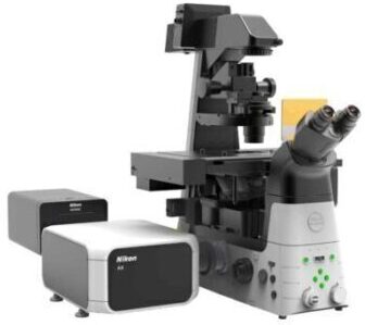
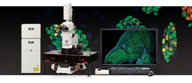
Confocal laser microscope system C2+/C2si+ Ready/C2si
The Nikon C2+ series offers the optimum confocal system to meet both your research and your budgetary needs.
C2+: Standard model boasts high resolution, high sensitivity, and high contrast. Suited for single laboratories or large research groups.
C2si+ Ready: Upgrade to C2si+ is possible by adding a spectral detector.
C2si+: Spectral confocal system featuring a 32-ch multianode spectral detector. A spectral bandwidth of 320 nm can be captured in a single scan.

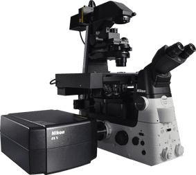
Configuration with spectral confocal system C2si+
[Note] Upgrade to the macro confocal microscope system is through a combination of the C2+ series and the AZ100 microscope. The AZ100M andA1+ series are not compatible.
Sample images captured with AZ-C1
| AZ100 optical zoom | 1-8x (zoom ratio 8 : 1) | |
|---|---|---|
| C2+ confocal scan zoom | 1x-1000x (continuous variable) | |
| Stand | AZ-STE EPI stand/AZ-STD DIA stand | Focus range: 85 mm stroke
Coarse focus: 18.5 mm/rotation Fine focus: 3.27 mm/rotation Stage focus: 10 mm range with 0.27 mm/rotation |
| Objective | AZ-Plan Apochromat 0.5x (NA 0.05/W.D. 54 mm), AZ-Plan Apochromat 1x (NA 0.1/W.D. 35 mm), AZ-Plan Fluor 2x (NA 0.2/W.D. 45 mm), AZ-Plan Apochromat 4x (NA 0.4/W.D. 20 mm), AZ-Plan Fluor 5x (NA 0.5/W.D. 15 mm) | |
| Laser light source | Laser wavelength options | 440 nm,488 nm Ar laser (457nm,477nm488nm,514nm), 543 nm/561nm/594nm 633nm/638nm/640nm |
| Maximum number | 4 | |
| Laser control options | AOM/AOTF/manual | |
| Laser shutter | Motorized mechanical shutter (each laser) | |
| Standard fluorescence detector | Number of channels | C2si+: 3 channels, C2si+ Ready: 3 channels, C2+: 2 channels/3 channels |
| Display mode | 160 x 160 to 2048 x 2048 pixels | |
| Scanning speed | Standard mode:2 fps (512 x 512 pixels, bi-direction) Zoom:1-1000x Fast mode:8 fps (512 x 512 pixels, bi-direction) Zoom:8-1000x |
|
| Spectral detector (C2si+) | Number of channels | 32 channels |
| 1st dichroic mirror | 20/80 beam splitter | |
| Corresponding wavelength | 400-750 nm | |
| Wavelength resolution | 2.5/5/10 nm (switchable) | |
| Minimum wavelength step | 0.25 nm | |
| Display mode | 160 x 160 to 1024 x 1024 pixels | |
| Scanning speed | Standard: 0.5 fps (512 x 512 pixels, 32 channel simultaneous recording ) | |
| Power | Confocal system (PC, monitor, C2+ controller, AOM controller): Approx. 830 W (single phase AC 115 V, 7.2 A / AC 230 V, 3.6 A, with earth)
(Does not include microscopes and lasers.) |
|

 Developmental Biology/Embryology
 Reproductive Medicine