The Back-Illuminated sCMOS Microscopy Camera You’ve Been Waiting For..
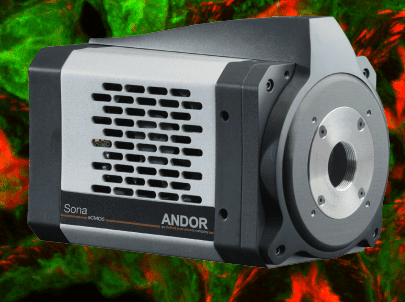
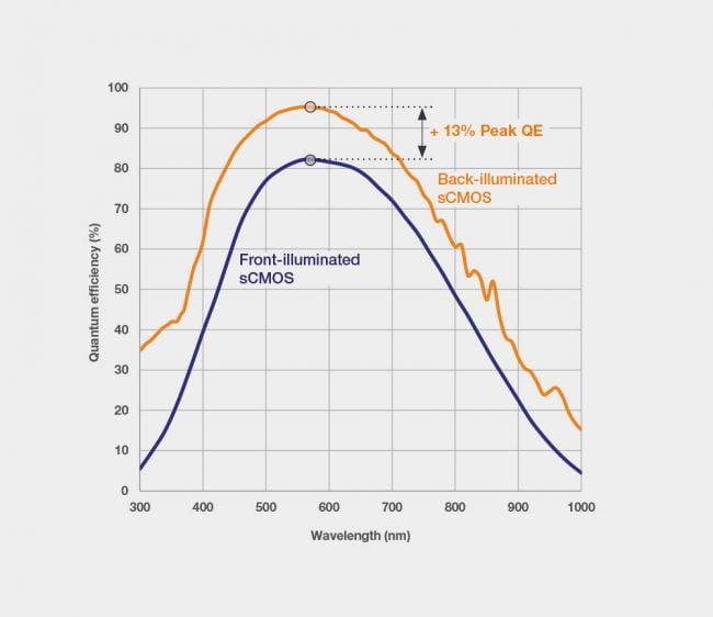
Sona is Andor’s latest high performance sCMOS camera platform, specifically for fluorescence microscopy. It launches with Sona 4.2B-11 and Sona 2.0B-11, each featuring 95% Quantum Efficiency (QE) and market-leading vacuum cooling to -45 °C.
Andor’s unique capability to deliver the ultimate in sCMOS sensitivity means signal to noise can be optimized in fluorescence microscopy under conditions of reduced excitation power, thus preserving living cells during extended measurement periods. The superior sensitivity of Sona also compliments reduction of fluorophore concentrations, thus minimally perturbing the cell’s physiology. Higher sensitivity means that exposure times can be shortened, facilitating faster frame rate measurements of dynamic processes, such as intracellular signalling mechanisms or cell motility. The ‘dual-amplifier’ approach to extended dynamic range is ideal for accurately imaging and quantifying challenging samples such as neurons. Furthermore, to achieve best-in-class quantification accuracy, Andor have implemented enhanced on-head intelligence to deliver market-leading linearity of > 99.7% across the whole dynamic range.
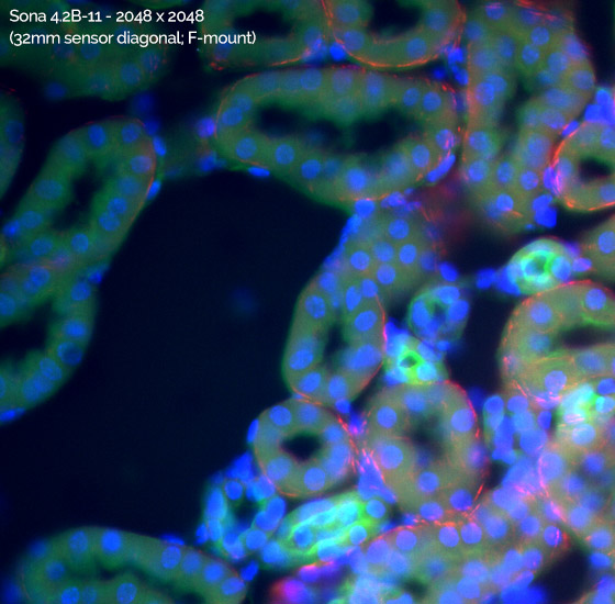
The flagship 4.2 Megapixel Sona 4.2B-11 model utilizes a unique technology approach that enables us to usefully access the entire 2048 x 2048 array, offering an impressive 32 mm sensor diagonal, harnessing the entire field of view available from the microscope. This is perfect for applications that require maximum information content, allowing large fields of cells, whole embryos or tissue samples to be captured with absolute clarity..

Sona 4.2B-11 Field of View Advantage: Sona 4.2B-11 with 2048 x 2048 array has a 62% larger field of viewthan a competing back-illuminated sCMOS camera with a 1608 x 1608 array. Captured using a Nikon Ti2 microscope with 60x objective and integrated 1.5x tube lens, accessing the full 2048 x 2048 array while preserving Nyquist resolving clarity. Note that the Andor Magnifying Coupler Unit (MCU) can also be use used to provide additional magnification across a wide range of microscope ports.
Sona is the only vacuum back-illuminated sCMOS platform. As well as affording superior minimization of the noise floor, the performance longevity benefits of Andor’s vacuum sensor enclosure should not be overlooked. Unless protected, back-illuminated silicon sensors are susceptible to attack from moisture, hydrocarbons and other gas contaminants, resulting in gradual performance decline, including QE decline. Andor’s proprietary UltraVacTM process uses a hermetic vacuum seal, completely preventing any gas and moisture ingress from the outside environment, avoiding moisture condensation on the sensor and the need to return to factory for repair.
95% QE & lowest noise – Prolonged live cell observations / measure accurate physiology
4.2 Megapixel & 32 mm F-mount (Sona 4.2B-11) – Capture maximum field of cells and large tissue samples
2.0 Megapixel and 22 mm C-mount (Sona 2.0B-11) – Ideal for modern microscopes that have C-mount ports up to 22 mm
Easily adaptable to 60x and 40x objectives – Combine with Magnifying Coupler Unit (MCU) – preserve optical clarity over a range of sample types.
Vacuum Cooled to -45 °C – Very weak signals require lowest noise floor: Don’t be limited by camera thermal noise!
The ONLY vacuum back-illuminated sCMOS – Andor’s proprietary UltraVac™ technology protects the sensor from (a) QE degradation, and (b) moisture condensation.
Anti-Glow Technology – Allows access to full 4.2 Megapixel array with long exposures – maximize field of view and sensitivity advantages.
48 fps (4.2 Megapixel); 70 fps (2.0 Megapixel) – Image highly dynamic samples without signal smear – e.g. cell motility, membrane dynamics, ion flux, blood flow.
Extended Dynamic Range mode – ‘One snap quantification’ across a 53,000:1 signal range – measure challenging samples such as neurons
> 99.7% linearity – Market leading quantitative accuracy over the whole signal range – confidence of measurement in any application where signal intensity indicates local concentration.
User configurable ROI – Adapt to a range of microscope port sizes. Push frame rates and save data storage space.
Fan and Water cooling as standard – Water cooling for maximum sensitivity and highly vibration sensitive set-ups, e.g. super-resolution and electrophysiology
USB 3.0 (USB 3.1 Gen 1) – A convenient high speed interface
| Key Specifications | |
| Sensor Type | GPixel 400 back-illuminated |
| QE Options | BV |
| Active Pixels | Sona 4.2B-11 – 2048 x 2048 Sona 2.0B-11 – 1400 x 1400 |
| Sensor Size | Sona 4.2B-11 – 22.5 mm x 22.5 mm (32mm diagonal) Sona 2.0B-11 – 15.5 mm x 15.5 mm (22 mm diagonal) |
| Pixel Size | 11 μm |
| Pixel readout rate | 200 MHz (12-bit mode) 100 MHz (16-bit mode) |
| Read Noise | 1.6 e- (median) |
| Maximum frame rate | Sona 4.2B-11 – 48 fps (12-bit); 24 fps (16-bit) Sona 2.0B-11 – 70 fps (12-bit); 35 fps (16-bit) |
| Maximum Quantum Efficiency | 95% |
| Dark current, e-/pixel/sec at -45 °C | 0.2 |
| Exposure modes | Rolling Shutter |
| Pixel well depth | 85,000 e- |
| Maximum dynamic range | 53,000:1 |
| Linearity | > 99.7% |
| Photon Response Non-Uniformity (PRNU) | < 0.5% |
| Data range | 12 bit (fastest frame rates) and 16 bit (max dynamic range) |
| Interface | USB 3.0 |