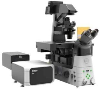
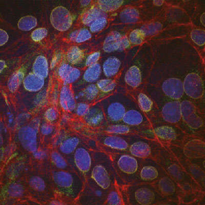
A confocal microscope that captures images with a 25 mm field of view, nearly twice the area of conventional point scanners.
Capturing images of large samples such as tissues, organs and live model organisms requires both extending the detectable area of cellular responses and increasing image capture speed. The A1 HD25/A1R HD25 confocal microscope has the largest field of view (25 mm) in its field, enabling users to expand the limits of scientific research.
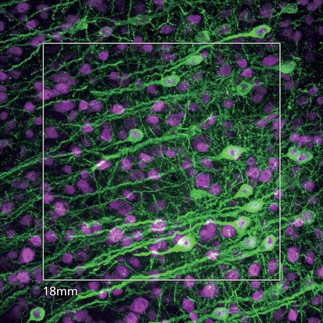
New 25mm FOV
When combined with the Ti2-E inverted microscope, the imaging area of the A1 HD25/A1R HD25 is nearly twice the conventional FOV of 18 mm, enabling the user to obtain significantly more data by capturing more of the specimen in each shot.
The large 25 mm FOV of the A1 HD25/A1R HD25 reduces both the required number of images for stitching large images and image acquisition times, enabling efficient, high-throughput imaging of even large-scale samples. The number of required images can be greatly reduced, in particular for large 3D (XYZ) image stitching.
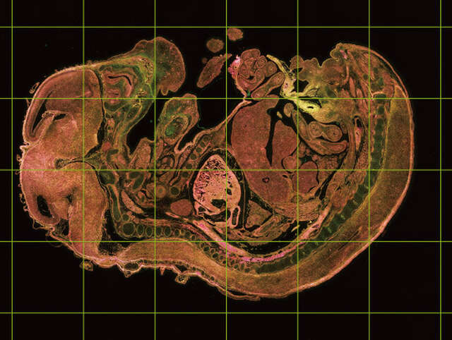 FOV 25 of A1 HD25/A1R HD25: a total of 24 frames |
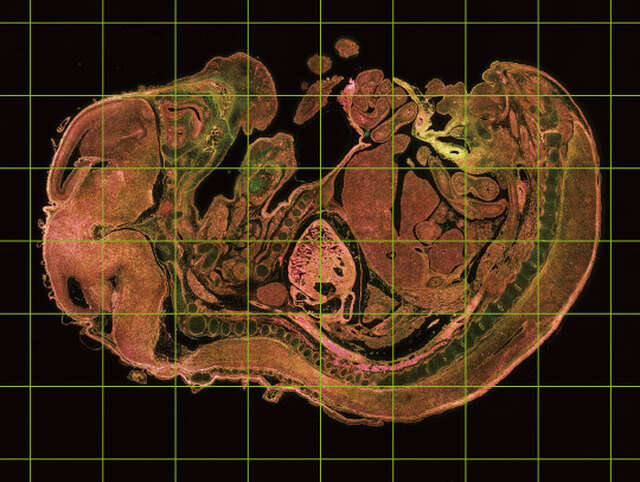 Conventional FOV 18: a total of 48 frames |
The combination of a high-speed resonant scanner and large field of view forms an ideal platform for high-resolution screening assays. It dramatically reduces the time needed to analyze multiple samples and conditions.
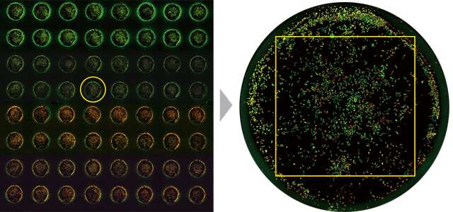 Left: A well in the 96-well plate is selected Right: The large FOV enables acquisition of entire wells (with a 4X objective) |
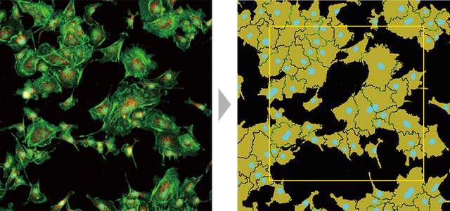 Large FOV enables measurement of larger areas and high-throughput analysis. |
1024 x 1024 pixels enables acquisition of high-resolution, high-quality images at lower magnifications, enabling compatibility with a wide range of samples.
 ① 1 x zoom (1024 x 1024 pixels)
|
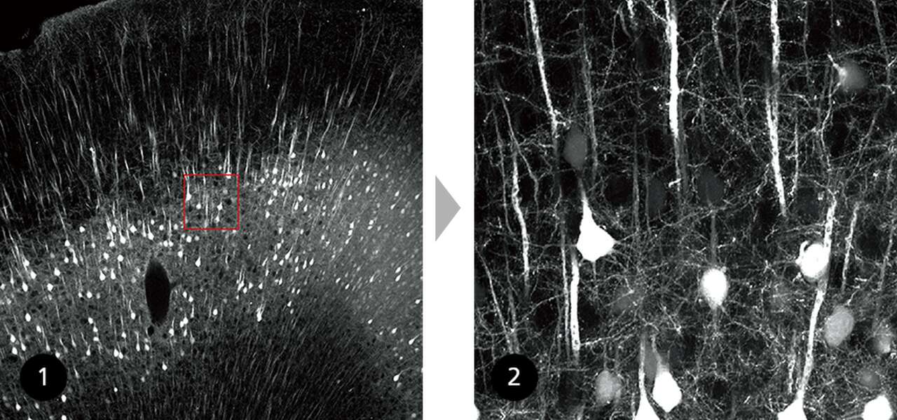
Comparison of a large FOV image and 6X zoomed image (1024 x 1024 pixels) of fine structures in a 2 mm brain slice of H-line mouse cleared with RapiClear1.52, SunJinLab.
Image courtesy of: Drs. Ryosuke Kawakami, Kohei Otomo, and Tomomi Nemoto, Research Institute for Electronic Science, Hokkaido University
High speed imaging capability up to 720 fps, in combination with a large field of view, dramatically increases imaging throughput. This scanning method reduces the exposure time of the sample to excitation light, minimizing phototoxicity and photobleaching.
Galvano scanner
 |
 |
 |
Resonant scanner
 |
 |
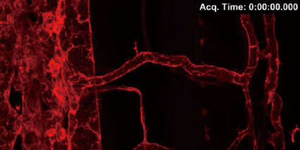 |
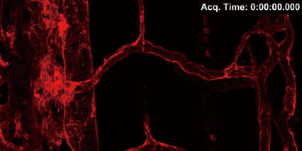
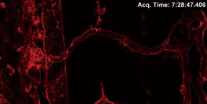
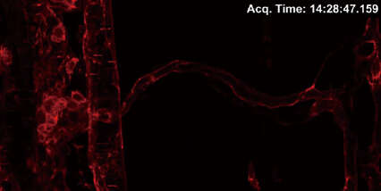
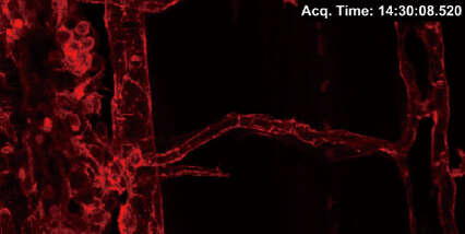
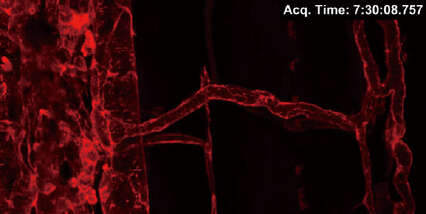
Comparison of photobleaching of fluorescent proteins when images are acquired using both galvano and resonant scanners. 3D time-lapse images of trunk vasculature in zebrafish larva expressing LIFEACT-mCherry (probe for F-actin) in endothelial cells were acquired every 30 minutes over a period of 15 hours using a galvano scanner (average of 2 images) and a resonant scanner (average of 64 images).
1024 x 512 pixels, 2X zoom, 100 Z-stack images
Note that photobleaching of LIFEACT-mCherry was dramatically suppressed using the resonant scanner.
Image courtesy of: Shinya Yuge Ph.D., and Shigetomo Fukuhara, Ph.D., Department of Molecular Pathophysiology, Institute of Advanced Medical Sciences, Nippon Medical School
Capture large-scale overview images as well as high magnification images with the same instrument. The 25mm FOV of the A1 HD25/A1R HD25 is effective for observation of large samples, while its 1Kx1K high-definition is ideal for the observation of minute structures.
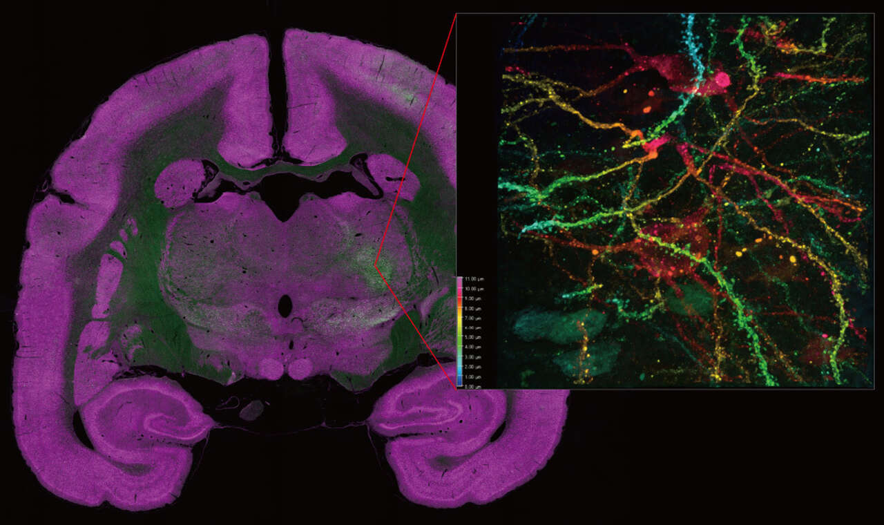
Stitched overview image of marmoset brain captured with CFI Plan Apochromat Lambda 10X objective and detailed image of dendritic spines captured with CFI SR HP Plan Apochromat Lambda S 100XC Sil objective
The A1-DUG-2 is a 4-channel detector unit equipped with high-sensitivity GaAsP PMTs, allowing acquisition of bright signals with minimal background noise, even when fluorescence is weak or the detector unit is used with a high-speed resonant scanner.
|
Images of HeLa cells labeled with MitoTracker, together with their intensity surface plots. Fluorescence intensities are indicated together with colors and heights in the 3D graph. There are major differences between GaAsP PMT (left) and Multi-Alkali PMT (right) in the background noise of and temporal change in the image. |
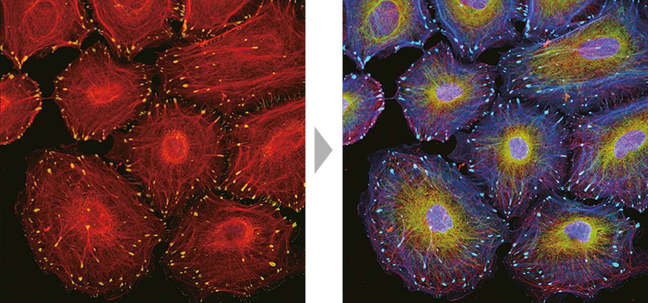
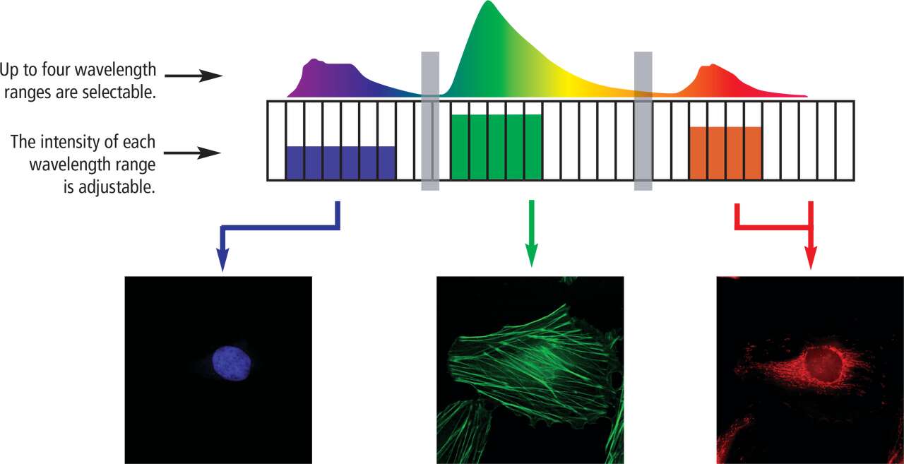
A1-DUVB-2 is equipped with a high-sensitivity GaAsP PMT and allows spectral imaging using both galvano and resonant scanners. This is a fully tunable emission detector unit capable of spectral imaging with user-defined emission bandwidths as low as 10 nm. Variable bandpass mode and continuous bandpass mode are selectable based on the applications and the images can be spectrally unmixed. The option to add a second fixed bandwidth emission channel enables simultaneous multi-channel imaging.
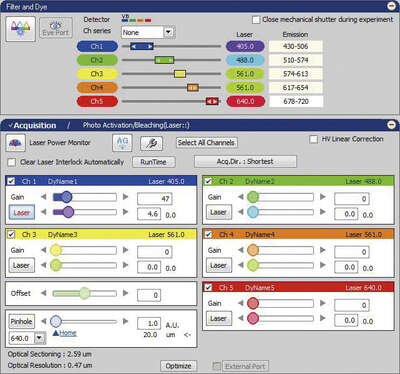 VB (Variable Bandpass) mode |
 VB (Variable Bandpass) mode allows maximum 5ch color image |
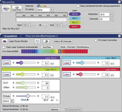 Unmixed Image CB (Continuous Bandpass) mode |
 CB (Continuous Bandpass) mode allows maximum 32 chspectrum imaging |
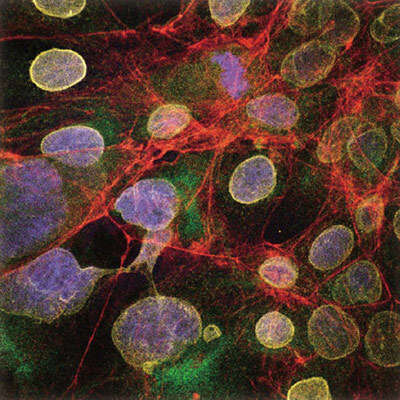
HeLa cells labeled with five-color fluorescence, Nucleus: DAPI, Vimentin: Alexa Fluor® 488, Lamin: Alexa Fluor® 568, Tubulin: Alexa Fluor® 594, Actin: Alexa Fluor® 633
Specimen courtesy of: Dr. Tadashi Karashima, Department of Dermatology, Kurume University School of Medicine
The A1-DUS can acquire spectral images at a high wavelength resolution of at least 2.5 nm. 32 channels of fluorescence spectra (up to a 320 nm wavelength range) can be acquired with a single scan, enabling fast imaging at up to 24 fps (512 x 32 pixels). Simultaneous excitation with up to four lasers enables spectral imaging across wider bands.
It precisely unmixes closely overlapping spectra of fluorescent labels and eliminates autofluorescence. Real-time unmixing during image acquisition is highly effective for FRET analysis.
Spectral and unmixed images of five-color-fluorescence-labeled HeLa cells
Specimen courtesy of: Dr. Tadashi Karashima, Department of Dermatology, Kurume University School of Medicine
The unique V-filtering function, with its capacity for filter-less intensity adjustment, allows arbitrary spectral ranges to be selected from 32 channels and combined to perform the filtering function of up to four filters, enabling imaging with the optimal intensity of each fluorescence probe.
Nikon provides a broad range of high-NA objectives with unrivaled optical quality to redefine the boundaries of confocal imaging. Options include silicone oil immersion objectives for thick live cell imaging, large-FOV low-magnification objectives and easy-to-use dry objectives. Chromatic aberrations are corrected from ultraviolet to near infrared range, enabling excellent multicolor imaging.
| A1 HD25 | A1R HD25 | ||
|---|---|---|---|
| Scan head input/output port | 1 laser input port 2 signal output ports for standard, spectral and optional detector*1 |
||
| Laser | LU-N3 3-laser unit | 405 nm, 488 nm, 561nm lasers are installed; built-in AOTF *Cannot be used with A1-DUS spectral detector |
|
| LU-N4/N4S 4-laser unit | 405 nm, 488 nm, 561 nm,640 nm lasers are installed; built-in AOTF *LU-N4 cannot be used with A1-DUS spectral detector |
||
| LU-NV series laser unit | Compatible lasers : 405 nm, 445 nm, 458 nm, 488 nm, 514 nm, 532 nm, 561 nm, 594 nm, 640 nm, 647 nm ; built-in AOTF | ||
| Standard fluorescence detector | Wavelength | 400-750 nm | |
| Detector | A1-DU4-2 4 Detector Unit: 4 Multi-Alkali PMTs A1-DUG-2 GaAsP Multi Detector Unit: 2 GaAsP PMTs + 2 Multi-Alkali PMTs |
||
| Filter cube | 6 filter cubes commonly used for a microscope mountable on each of three filter wheels Recommended wavelengths: 450/50, 482/35, 515/30, 525/50, 540/30, 550/49, 585/65, 595/50, 700/75 |
||
| Diascopic detector (Option) | Wavelength | 485-650 nm | |
| Detector | Multi-Alkali PMT | ||
| FOV | Ti2-E:Square inscribed in a ø25 mm circle Ni-E/FN1:Square inscribed in a ø18 mm circle |
||
| Image bit depth | 4096 gray intensity levels (12 bit) | ||
| Scan head | Standard image acquisition | Scanner: galvano scanner x2 Pixel size: max. 4096 x 4096 pixels Scanning speed: Standard mode: 1.4 fps (512 x 512 pixels, bi-direction, 0.72x zoom), 2 fps (512 x 512 pixels, bi-direction, 1x zoom) Fast mode: 10 fps (512 x 512 pixels, bi-direction, 8x zoom), 200 fps (512 x 16 pixels, bi-direction, 8x zoom)*2 Zoom: 1-1000x continuously variable Scan mode: X-Y, X-T, X-Z, XY rotation, Free line, Line-Z |
|
| High-speed image acquisition | — | Scanner: resonant scanner (X-axis, resonance frequency 7.8 kHz), galvano scanner (Y-axis) Pixel size: max. 1024 x 1024 pixels Scanning speed: 15 fps (1024 x 1024 pixels), 30 fps (512 x 512 pixels), 60 fps (256 x 256 pixels) to 720 fps (512 x 16 pixels), 7,800 lines/sec (line speed) Zoom:0.72x, 0.82x, 0.9x, 1x, 1.2x, 1.5x, 1.75x, 2x, 2.4x, 3x, 4x, 5x, 6x, 7x, 8x Scan mode: X-Y, X-T, X-Z Acquisition method: High-speed image acquisition |
|
| Dichroic mirror | Low-angle incidence method, Number of positions: 8 Standard filter: 405/488/561/640, BS20/80 Optional filter:405/488, 405/488/561, 405/488/543/640, 457/514 |
||
| Pinhole | 12-256 μm variable (1st image plane) | ||
| Spectral detector (option) | A1-DUS spectral detector unit | Number of channels: 32 Wavelength detection range: 400 – 750 nm Spectral image acquisition speed: 4 fps (256 x 256 pixels) Maximum pixel size: 2048 x 2048 (Spectral mode/Virtual filter mode) Wavelength resolution: 2.5/6.0/10.0 nm, wavelength range variable in 0.25 nm steps Compatible with galvano scanner only |
|
| A1-DUVB-2 GaAsP detector unit | Number of channels: 1 GaAsP PMT with variable emission plus 1 optional GaAsP PMT (A1-DUVB-OP) with a user-defined dichroic mirror and barrier filter Wavelength detection range: 400 – 720 nm, narrowest: 10 nm, broadest:320 nm Maximum pixel size: 4096 x 4096 (CB mode/VB mode) Wavelength resolution: 10 nm, wavelength range variable in 1 nm steps Compatible with galvano and resonant scanners |
||
| Z step | Ti2-E: 0.01 μm, 0.02 μm (with encoder control), FN1 stepping motor: 0.05 μm, Ni-E: 0.025 μm | ||
| Compatible microscopes | ECLIPSE Ti2-E inverted microscope, ECLIPSE FN1 fixed stage microscope, ECLIPSE Ni-E upright microscope (focusing nosepiece type and focusing stage type) |
||
| Option | Motorized XYZ | Motorized XY stage (for Ti2-E/Ni-E), High-speed Z stage (for Ti2-E), High-speed piezo objective-positioning system (for FN1/Ni-E) | |
| Photostimulation module*3 (for Ti2-E) |
XY galvano scanning unit (Light source: LU-N3/N4 Laser Unit) DMD module (Light source: C-LEDFI Epi-FL LED illuminator, LU-N3/N4 laser unit) Stimulation form: ROI/line/point Stimulation mode: Sequential, Simultaneous |
||
| Software | Acquisition/analysis | Basic software: NIS-Elements C Optional software for high-resolution acquisition: NIS-Elements C-ER |
|
| Display/image generation | 2D analysis, 3D volume rendering/orthogonal, 4D analysis, spectral unmixing | ||
| Image format | JP2, JPG, TIFF, BMP, GIF, PNG, ND2, JFF, JTF, AVI, ICS/IDS | ||
| Application | FRAP, FLIP, FRET(option), photoactivation, three-dimensional time-lapse imaging, multipoint time-lapse imaging, colocalization | ||
| Control computer | OS | Windows 10 Pro 64bit, English or Japanese version, OS Version 1709 Windows 7 Professional, 64bit, SP1 English or Japanese version, Windows Update KB3118401 or later |
|
| CPU | Intel Xeon W-2125 (4.0GHz, quad core, 8.25 MB, 2666 MHz) or higher | ||
| RAM | 32GB or 64GB | ||
| HDD | 1st HP Z Turbo G2 512GB PCIe M.2 SSD 2nd SATA HDD 2TB |
||
| Optical Drive | Super Multi drive, up to x16 speed or higher | ||
| Graphics | NVIDIA Quadro P600 or higher (NIS-Elements C-ER: NVDIA Quadro P4000) (PCI Express/two-screen split display supported) |
||
| Extension slot | Two PCI Express 3.0 (x16) slots (one slot to be used for graphics) One PCI Express 3.0 (x8) slot Two PCI Express 2.0 (x4) slot |
||
| LAN port | 10/100/1000 Network/Interface x 2 (for connection to controller, for connection to external LAN) | ||
| Monitor | 1600 x 1200 or higher resolution, dual monitor configuration recommended | ||
| Recommended installation conditions | Temperature 23 ± 5 ºC, humidity 70 % (RH) or less (non-condensing) | ||
*1 FCS/FCCS/FLIM is possible in combination with third-party systems
*2 Fast mode is compatible with zoom 8-1000x and scanning modes X-Y and X-T. It is not compatible with Rotation, Free line, CROP, ROI, Spectral imaging, Stimulation and FLIM.
*3 Adapter for Ti2-LAPP system, light source, hybrid dichroic mirror for photoactivation and imaging, and control board are all required.
| Scan Head | 277(W) x 163(H) x 366(D) mm | Approx. 14.5 kg |
|---|---|---|
| Controller | 360(W) x 575(H) x 599(D) mm | Approx. 40 kg |
| A1-DU4-2 4 Detector Unit | 360(W) x 199(H) x 593.5(D) mm | Approx. 16 kg |
| A1-DUG-2 GaAsP Multi Detector Unit | 360(W) x 199(H) x 593.5(D) mm | Approx. 16 kg |
| A1-DUS Spectral Detector Unit | 360(W) x 323(H) x 593.5(D) mm | Approx. 26 kg |
| A1-DUVB-2 GaAsP Detector Unit | 360(W) x 114(H) x 595.5(D) mm | Approx. 10 kg |
| LU-N4/N4S/N3 Laser Unit | 360(W) x 210(H) x 593.5(D) mm | Approx. 19 kg |
| LU-NV Laser Unit | 400(W) x 781(H) x 685(D) mm | Approx. 70 kg |
| LU Controller Box B (for LU-NV) | 400(W) x 123(H) x 687(D) mm | Approx. 7 kg |
| A1 HD25/ A1R HD25 | Scan Head and Controller | Input 100-240V ± 10%, 50-60Hz, 5A-2A |
|---|---|---|
| Computer Unit | Input 100-240V ± 10%, 50-60Hz, 12A-10A | |
| Laser Unit | LU-N4/LU-N4S/LU-N3 | Input 100-240V ± 10%, 50-60Hz, 2A max. |
| LU-NV Series | Input 100-240V ± 10%, 50-60Hz, 4.8A max. | |
| LU Controller Box B (for LU-NV) | Input 100-240V ± 10%, 50-60Hz, 1A max. | |
| Microscope | Inverted Microscope Ti2-E and HG Fiber Illuminator Intensilight | Input 100-240V ± 10%, 50-60Hz, 6.3A max. |
Note: When an air compressor is used with a vibration isolated table, an additional power source is necessary.