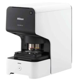
Research Microscope Power in a Benchtop Assay Instrument
Simple Operation
Minimal experimental setup and complexity by AI-driven assays and analysis.
Easy Cellular Imaging
No need to master complicated microscope hardware and software – ECLIPSE Ji makes data collection and cellular imaging assays easy.
High Quality Optics
Renowned Nikon optics provide clear and sharp images on plate assay devices.
Smart Experiments with Automated Assays
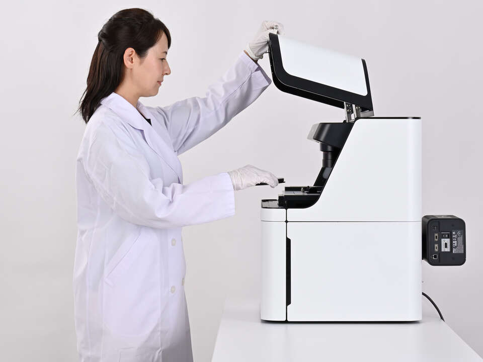
ECLIPSE Ji fits standard laboratory benchtops, has built-in vibration dampening hardware, and shields the sample from bright room light. There is no need for dedicated space or dark rooms to perform efficient imaging assays.
Utilizing Nikon’s precision optical hardware, all of the advantages of high sensitivity and resolution from a research-level microscope are retained by an AI-driven, easy-to-operate benchtop laboratory instrument.
Preconfigured and optimized turnkey assay experiments minimize time spent defining parameters, maximizing data collection.
Intensity MeasurementCompares protein expression level changes in cells and cell nuclei in multiple wells. |
Cell Counting (endpoint)Counts the number of cell nuclei in a fixed sample and the area of the well occupied by cells. |
Transfection EfficiencyInvestigates the percentage of cells expressing the target protein, and reports the expression efficiency. |
Size & Morphological analysisAnalyzes cellular morphology with measurements of the cell nucleus, cytoplasm, and the size of the cell region. |
CytotoxicityMeasures the percentage of dead cells among all cells and evaluates cytotoxicity. |
|
|
Nuclear TranslocationMeasures the nuclear translocation of NF-κB, indicating an extra-cellular stimulus. |
AutophagyMeasures the number of autophagosomes, their area, and their fluorescence intensity. |
PhagocytosisMeasures the number, area, and fluorescence intensity of bioparticles taken into the cell by phagocytosis. |
EndocytosisMeasures the number, area, and fluorescence intensity of granules formed by endocytosis, which are taken up from outside the cell. |
Micronucleus testMeasures the number of cells containing micronuclei or multiple nuclei. |
Mitochondrial toxicityMeasures the number, area, and fluorescence intensity of mitochondria. |
Neurite OutgrowthMeasure the number and length of neurites protruding from neuron cell bodies. |
Wound HealingMeasure the recovery of filled area in an artificially-created wound over time. |
Cell Counting (proliferation)Measure changes in confluency and count nuclei in live samples over time. |
ECLIPSE Ji’s Smart Experiment software interface uses newly developed artificial intelligence (AI), implemented to minimize errors and maximize data collection.

Single-Cell Imaging and Analysis with Ease
AI based on Deep Learning defines acquisition settings and image analysis parameters, saving researchers valuable time at the microscope.
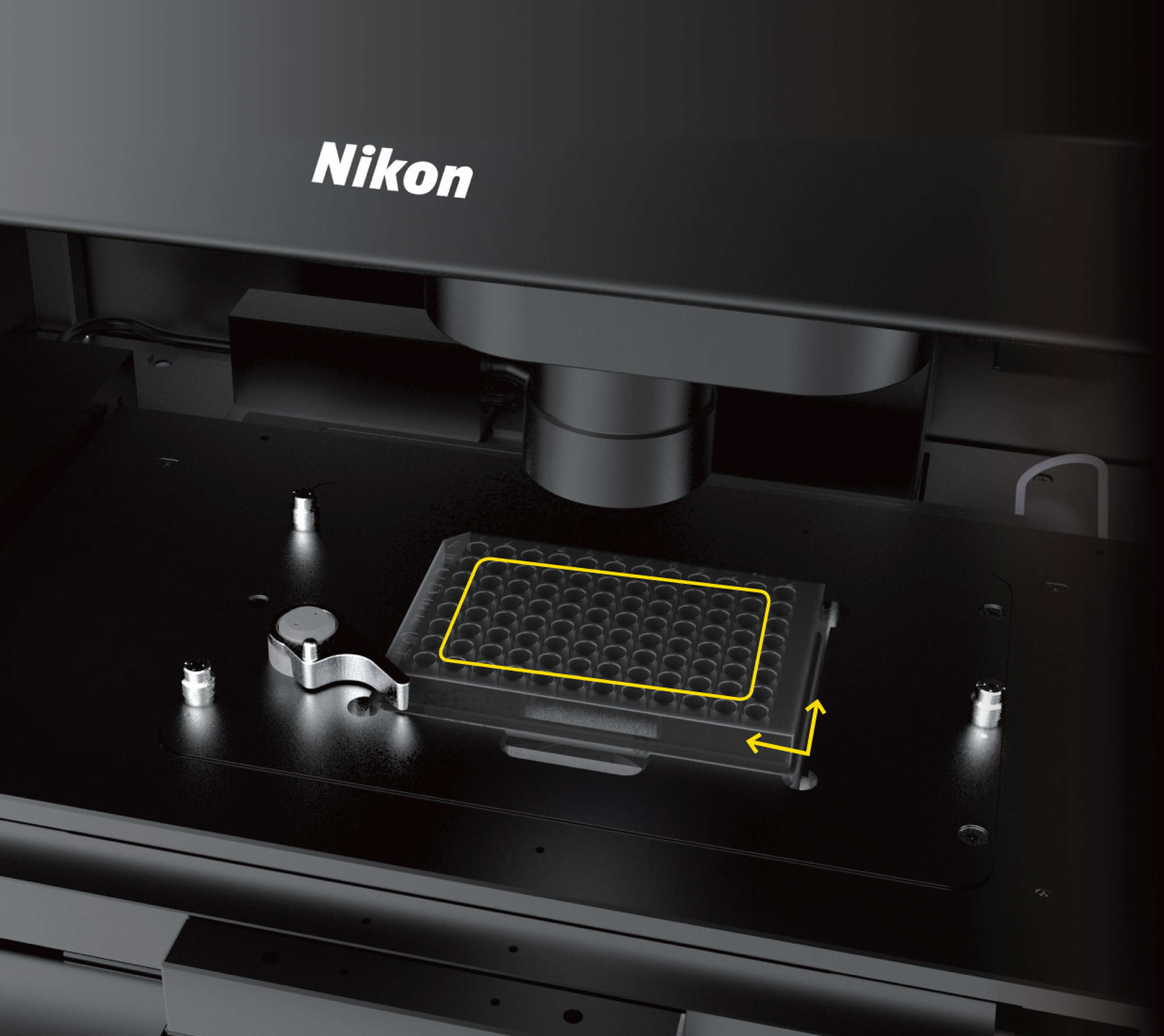
Plate type and dimensions are automatically detected. There is no need to select from lists or manually enter plate data.
A rapid preview across the entire plate determines which wells have sample present, allowing users to easily skip empty wells.
There is no need for troublesome tuning of light intensity and exposure time. The optimal exposure settings for image analysis are automatically calculated from the luminance values of all wells.
No alignment work is required. The system automatically detects and corrects the plate position.
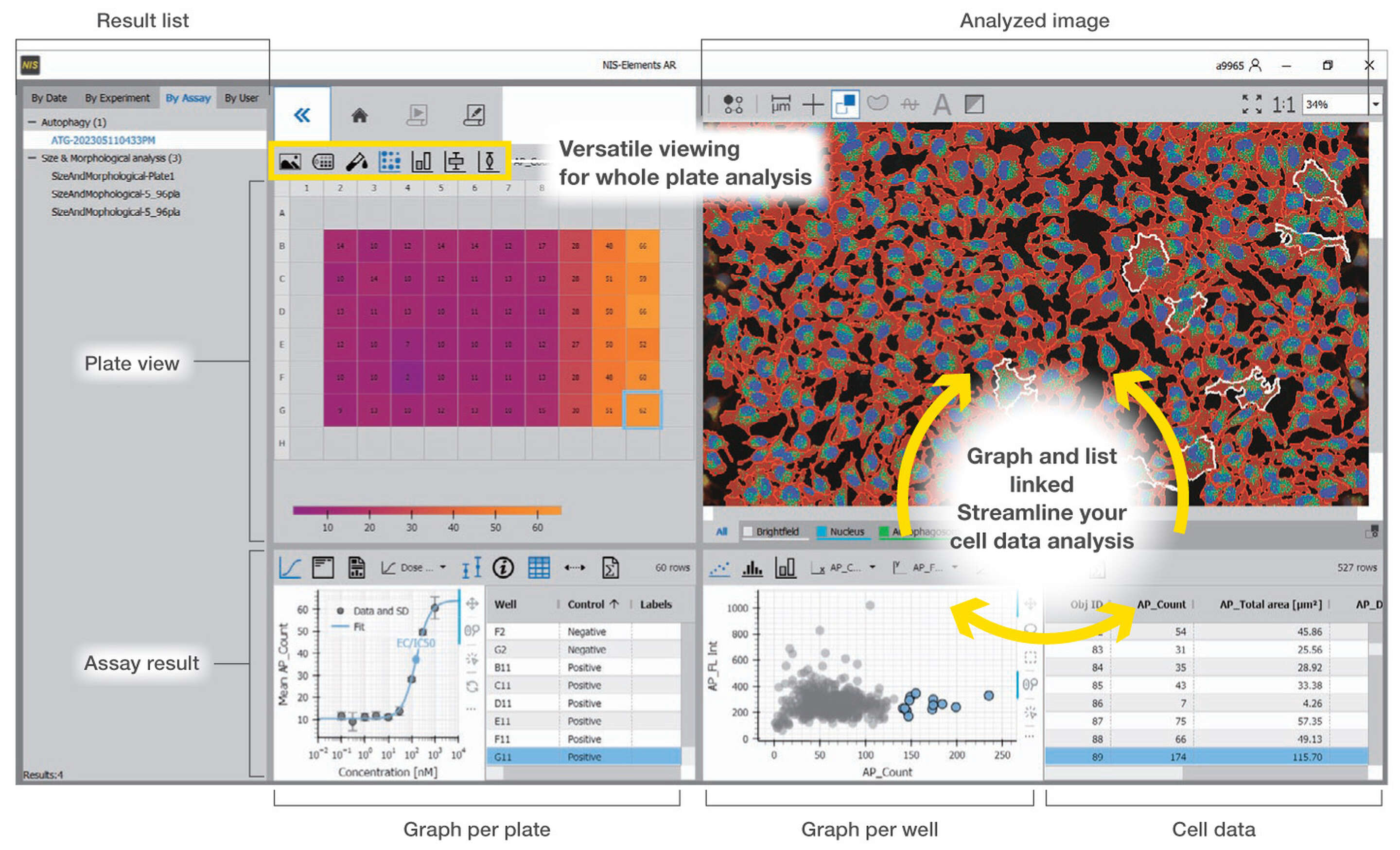
User Interface Designed for Data-Rich Microscopy
Images and corresponding analysis data for the plate, well, and each cell is contained in an interactive and linked interface. Users can navigate and quickly visualize trends and results.
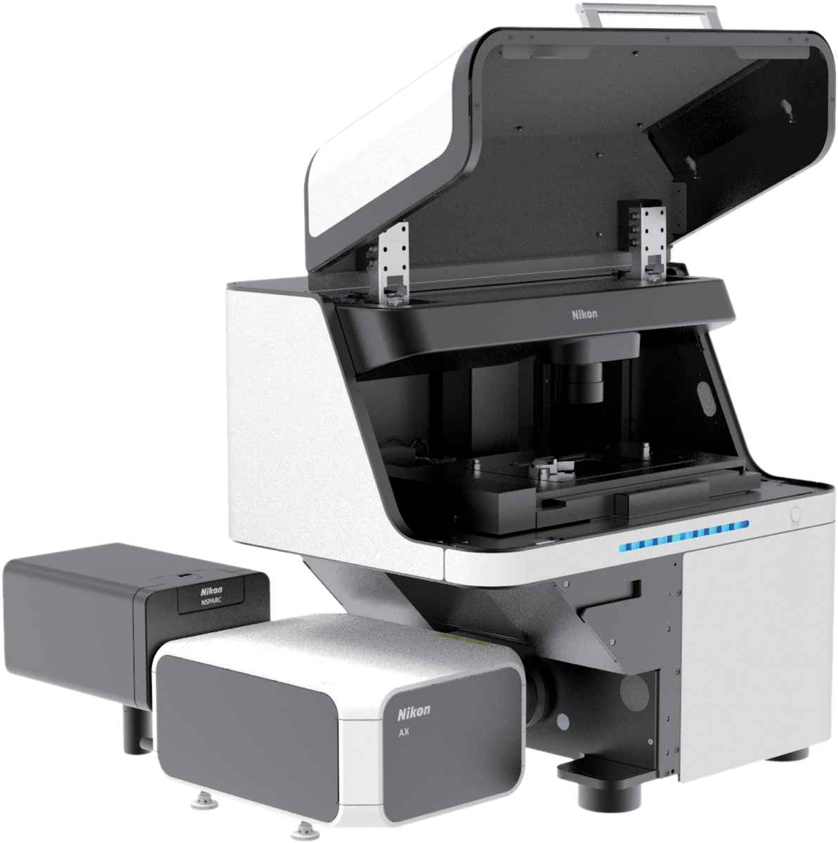
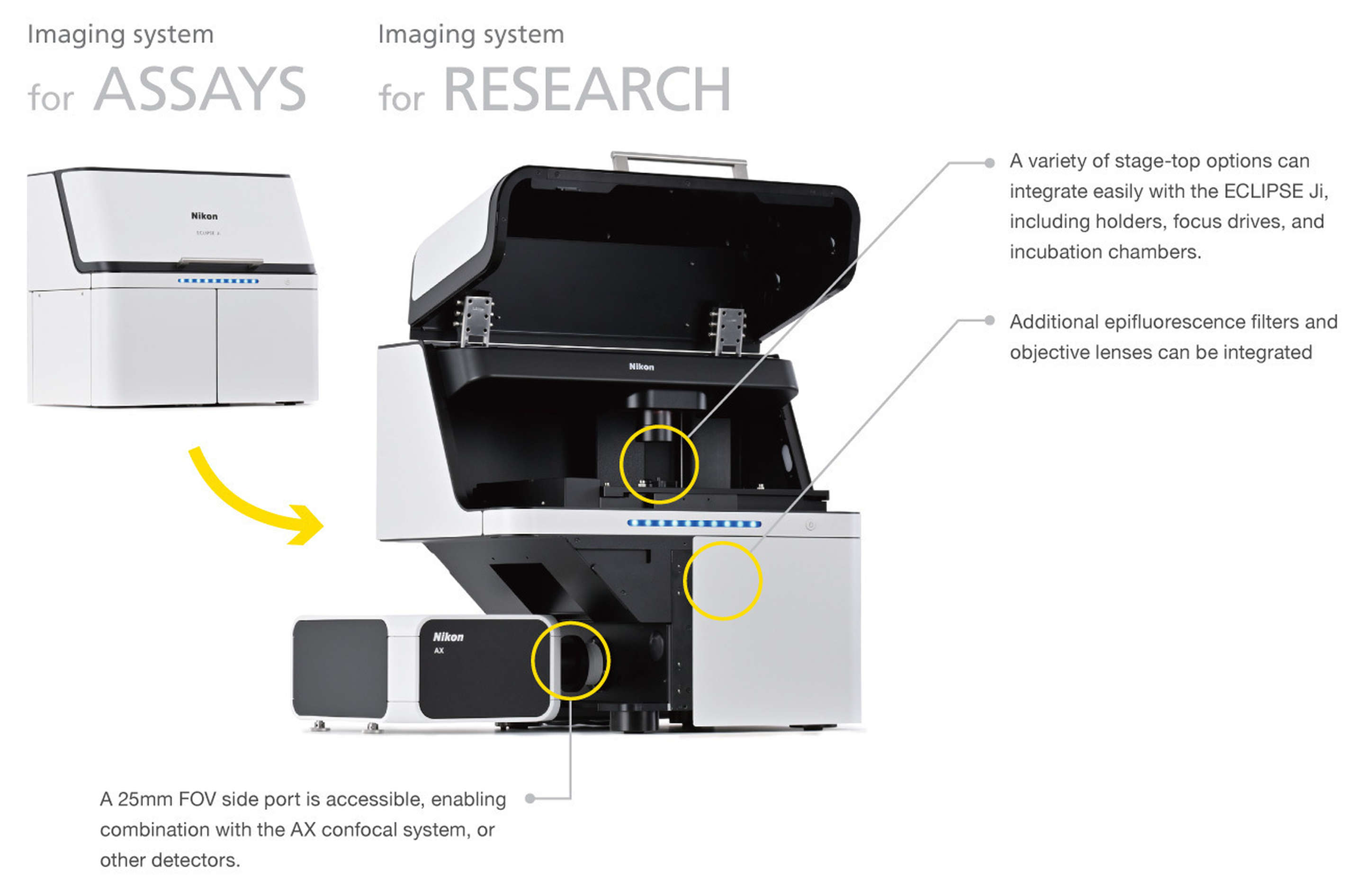
ECLIPSE Ji: a Multi-Role Digital Inverted Microscope
Outside of plate assays, ECLIPSE Ji can also serve as a digital research microscope, and can be integrated with a variety of peripheral hardware ranging from filter wheels through confocal systems such as “AX“, or high-sensitivity cameras.
*The design and specifications may differ from the actual product.
| ECLIPSE Ji | |
|---|---|
| Observation methods | Brightfield, Epi-fluorescence |
| Optical system | CFI Infinity Optical System Observation Optical System: Inverted image observation |
FOV 25
Optical path switching: Switching between the built-in camera optical system and the left side portBuilt-in cameraImaging device: 7.8 megapixel monochrome CMOS sensor
Output signal Tone: Monochrome 12 bits/8 bits
Frame rate: Maximum 18 fps
Output pixel number: 2800×2800 pixels (when assay used)FocusingDrive system: Motorized (Via PFS nosepiece objective lens up/down movement)Focusing stroke: About 10 mm
Focusing speed: Maximum driving speed 2.5 mm/secPFS*Focal point maintenance control: Infrared light projecting method
Applicable observation methods:
Brightfield, Fluorescence observationTransmission illumination sectionKoehler illumination Light source: LEDStageStroke: X: ±59 mm, Y: ±39.5 mm
Maximum drive speed About 25 mm/secNosepieceObjective lens mounting holes: 6
Nosepiece drive method: MotorizedFluorescence cube turretNumber of filter cubes that can be mounted: 6
(Compatible with wide-field filter cubes)
Turret
drive method: MotorizedLight distribution sectionLight source used: D-LEDI2 fluoresce LED light sourcePC interfaceUSB interface: Device interface (for built-in camera)
B connector
USB 3.0 (SuperSpeed)Input rating100V-240VAC±10%, 3.0 A, 50/60 HzPower consumption320 WPower source cord- 100 to 120 V: Power source cord of 3 conductor grounding Type SVT, NO.18 AWG, 3 m long maximum, rated at 125VAC minimum with detachable receptacles conforming to UL specifications
– 220 to 240 V: Power source cord of 3 conductor grounding Type H05VV-F 1 mm2, 3 m long maximum, rated at 250VAC minimum with detachable receptacles conforming to EU/EN specifications
*PFS: a function that automatically corrects focal point displacement due to the passage of time and/or stage movement.
The Open Architecture Digital Inverted MicroscopeThe ECLIPSE Ji (Ji) is Nikon’s first all-digital research grade inverted microscope. With no eyepieces, this microscope is designed to be easy to learn and use, while maintaining the optimum optical quality and large field of view (FOV) Nikon microscopes are well-known for.Additionally, Nikon’s 4th generation perfect focus system (PFS) is integrated into Ji for reliable long-term observation of specimens.Ji’s integrated enclosure means users can navigate their samples in brightly-lit environments or even remotely, using the embedded scientific grade CMOS detector, or use any number of other possible detector options, depending on the research application.