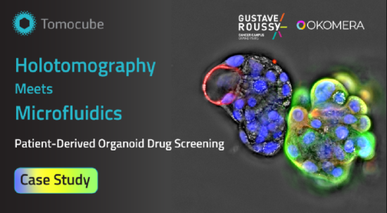
Welcome to the latest edition of our newsletter, featuring a new Case Study conducted in collaboration with Okomera and the research team from Institut Gustave Roussy.
This study highlights how Tomocube’s holotomography technology, when integrated with Okomera’s droplet-based microfluidics, enables detailed, label-free analysis of chemotherapy response in patient-derived organoids (PDOs). Our approach provides quantitative insights into the complex architecture of heterotypic PDOs, distinguishing tumor–stroma organization and microenvironmental responses.

|
Okomera’s droplet-based organoid culture combined with Tomocube holotomography and TomoAnalysis enables label-free, quantitative monitoring of drug responses in patient-derived organoid models. |
🔬 Highlights from the study:
Drug sensitivity profiling: Okomera’s microfluidic platform enabled a reliable assessment of carboplatin sensitivity in Luminal B PDOs using only a minimal sample from a patient biopsy.
Heterotypic PDO characterization: Correlative imaging of HT and immunofluorescence reveals distinct cellular architectures, with both tumor cells and stromal-like compartments.
Tumor vs. stroma distinction: HT identified stromal compartments with higher refractive index (RI) granularity than tumor regions, suggesting a potential label-free marker of stromal presence.
Stromal modulation of drug response: Stroma-rich organoids displayed higher resistance against carboplatin treatment, consistent with the role of the tumor microenvironment in mediating therapy resistance.
These results highlight how combining microfluidics and HT provides a deeper insight into drug response mechanisms beyond conventional endpoint assays.

Correlative imaging reveals tumor–stroma heterogeneity in biopsy-derived organoids. Immunostaining revealed heterogeneous tumor cell populations and stromal-like regions lacking epithelial markers (upper panel). HT further distinguished compartments, showing higher granularity in stromal-enriched regions (CK5/CK8-, pink box in lower panel) than in tumor-enriched regions (CK5/CK8+, blue box).
Tomocube holotomography: label-free 3D imaging and analysis
The HT-X1 series expands organoid research by providing universal, label-free imaging and quantitative analysis applicable to any PDO culture, revealing biophysical dynamics invisible to conventional assays.
By reconstructing 3D RI tomograms of live organoids without staining, it delivers:
Non-invasive, long-term imaging of optically thick organoids in flexible experimental design.
Quantitative biophysical readouts, such as dry mass, protein concentration, compactness, and granularity, for direct characterization of tumor-stroma heterogeneity and cell states.
AI-powered TomoAnalysis pipelines for reproducible segmentation and quantification—extensible to diverse organoid models and treatment studies.
Okomera microfluidics: high-throughput organoid culture
Okomera’s droplet-based microfluidic platform enables rapid, biopsy-ready PDO generation from as few as 30 cells. Within minutes, cells are encapsulated into droplets that form uniform organoids, enabling:
High-throughput multiplexed screening with barcoded drug libraries (O-Plex).
Fast turnaround, with compact 3D organoids formed within 48 hours.
Automated AI-driven analysis of growth, viability, and dose–response curves.
This streamlined approach allows researchers to maximize limited patient material while testing a wide range of drug conditions in parallel.
By integrating Tomocube’s HT and Okomera’s microfluidic technology, researchers can combine high-throughput screening with deep, label-free imaging—uncovering drug resistance and tumor heterogeneity that conventional methods often miss.