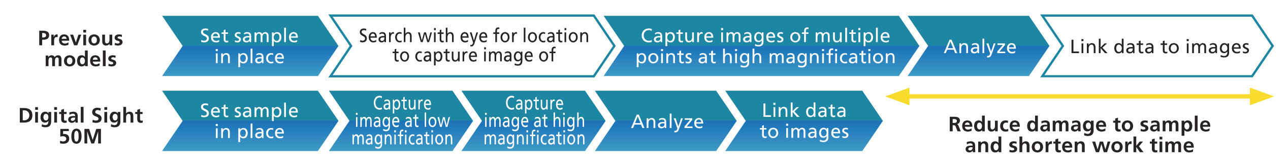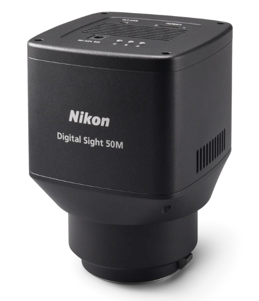
The monochrome digital camera Digital Sight 50M for microscopes is optimized to increase workflow efficiency. In addition to its large number of pixels, large field of view, and speed, it comes with dedicated software that makes it effective for screening large volumes of samples. It is perfect for not only academic research but also drug discovery.
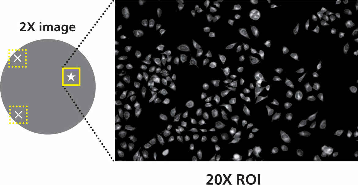
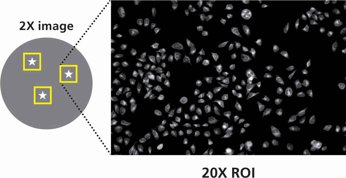
With an actual field of view of 7 mm when using a 2X objective lens, it is possible to capture single-shot images of wide areas. You can also quickly check both the overall image of large volumes of samples, such as in well plates, and regions of interest of a sample, which increases reproducibility of experiments.
The improved Digital Sight 50M boasts 3.8 times the number of pixels and 2.5 times the resolution of previous models. Even when using a low-magnification, high-NA objective lens, it fully demonstrates optical capabilities. It is also possible to obtain highly reliable data of small regions when analyzing images.
 |
 |
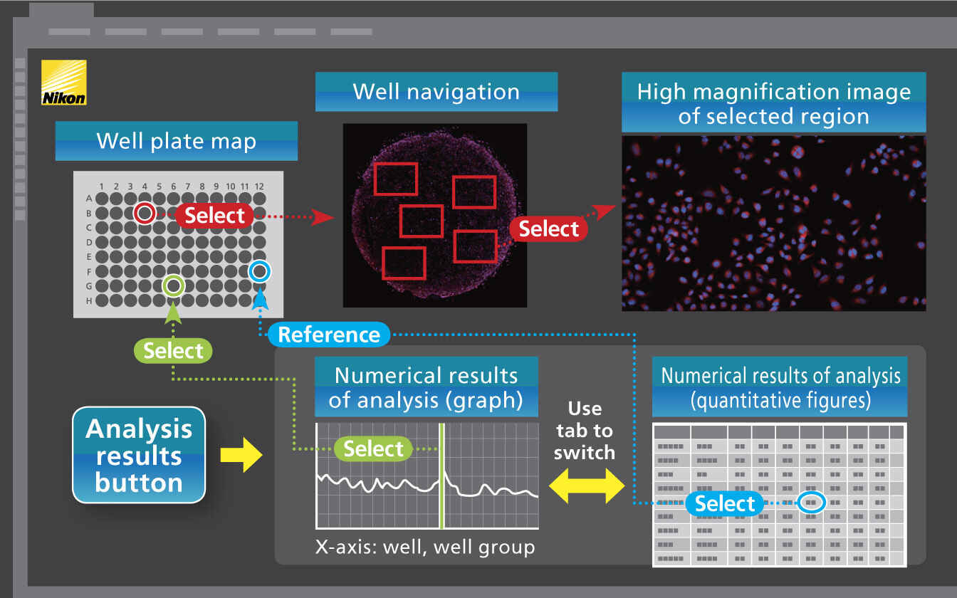
The Digital Sight 50M can be integrated with NIS-Elements HC software to support pre- and post-capture analysis. It is possible to set up a flow from well selection, automatic detection of image ROI, and displaying of analysis results.
 |
 |
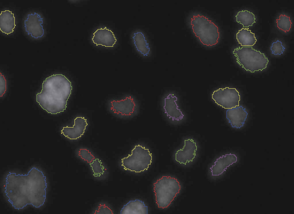
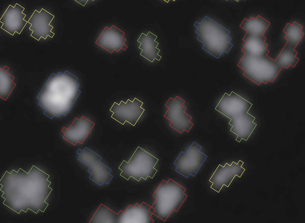
Detects even faint fluorescent signals
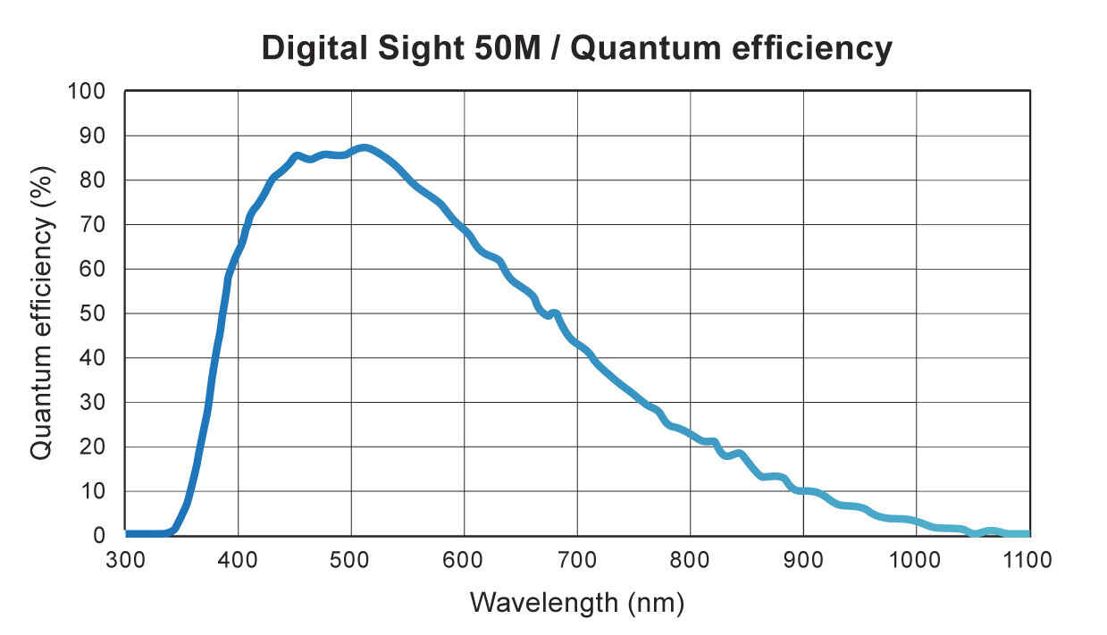
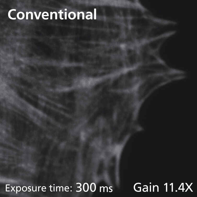
The Digital Sight 50M combines high spatial resolution with a 3.76 μm pixel size and achieves high quantum efficiency, peaking at 85%, which allows for even dim samples to be detected.
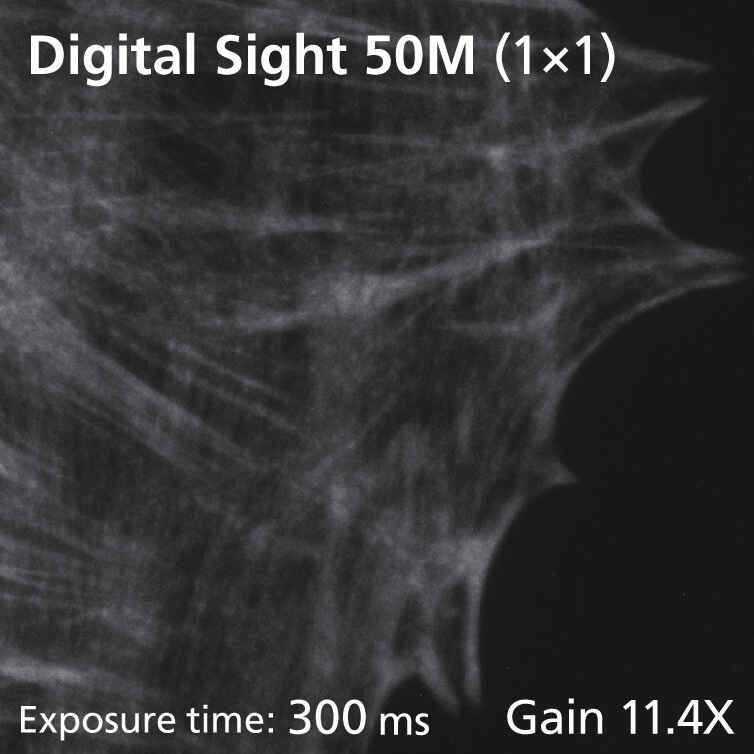
Acquires dim fluorescent signals with ultra-low noise
Both the 6e-read noise coupled with a large full-well capacity and 1.0e-/p/s dark current allow the acquisition of 14-bit fluorescence images with very little noise.
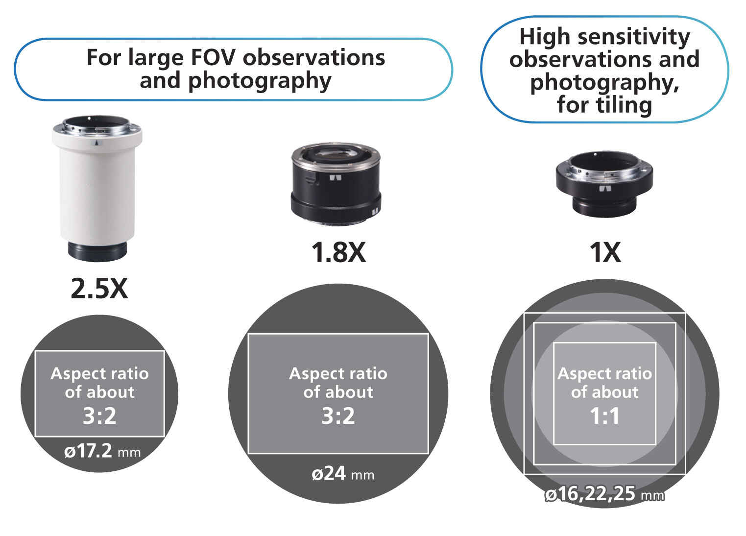
2.5X, 1.8X, and 1X adapters are available for different uses
The Digital Sight 50M offers the large CIS (Nikon FX format) that makes wide field-of-view (FOV25) observations possible. There are three adapters for different uses: 2.5X and 1.8X adapters for high-resolution single shots of 60 megapixel; and a 1X adapter for samples that require high sensitivity and low noise, such as image tiling.
Fluorescence time-lapse imaging through the integration with NIS-Elements software
Large field of view, high pixel density, and low noise make the Digital Sight 50M ideal for time-resolved imaging applications.
Flexible balance of quality and speed
There are three binning operation modes, making it possible to select the required speed and image quality. Maximum frame rate of 225.9 fps for high-speed imaging.
Fast focusing, even with fluorescence images
A high-sensitivity CMOS sensor and high-speed data transfer using USB 3.2 Gen 1 are combined to achieve 6 fps at the maximum number of pixels (60 megapixels) or a maximum speed of 27 fps (6.7 megapixels). This makes it possible to quickly focus on samples.
Capture regions of interest with much higher speeds of acquisition
Designate a region of interest within the camera field of view and then capture images of that desired region at high speed.
Nikon uses the NIS-Elements series as control software. NIS-Elements allows functions from basic imaging to control of the microscope and peripheral devices to be performed, as well as the measurement, analysis, and management of acquired images. Four basic packages and a variety of optional modules are available to suit every application and objective.
 Free packageThe bundled free package offers functions for the display of scale on live images, full-screen display, and more. The simple operation screen makes shooting easy. |
 Documentation packageThe documentation package is equipped with measurement and report creation functions. It enables general microscopic image acquisition in fields from biomedical to industrial, and is expandable through optional added features such as EDF and databases. |
  Research packageThe research package enables the construction of advanced image acquisition systems, including multidimensional imaging (up to 4 dimensions for Br, 6 dimensions for Ar), through integration with systemized microscopes. Sets equipped with a rich range of image processing and analysis functions are available for every application. |
Compatible OS: Windows® 10 and 11 64-bit Professional
| Digital Sight 50M | |
|---|---|
| Image sensor | Nikon FX-Format Monochrome CMOS image sensor Size: 35.8 × 23.8mm |
| Recordable pixels | All pixels: 9552 × 6336 |
| Lens mount | F-mount |
| Cooling method | Electronic cooling |
| ISO sensitivity (recommended exposure index) |
Equivalent to ISO 200 |
| Quantum efficiency | 85% at 500 nm |
| Full well capacity | 45000e- (typ.) |
| Readout noise | 6e– |
| Dark current | 1.0e-/p/s (Ta=25°C)(typ.) |
| Live display mode* (maximum fps) |
All pixels (9552 × 6336): 6 fps 3 x 3 pixels average @ 8 bit(ROI 640 × 480): 225.9 fps** |
| Exposure time | 150 μsec–120 sec |
| Photometry mode | Average photometry: Average intensity within the photometry area Peak photometry: Maximum intensity within the photometry area |
| Exposure control | One-time automatic exposure: Exposure time is adjusted automatically for one-time within the optimum range for the camera Continuous automatic exposure: Automatic exposure adjustment is performed continuously to keep the exposure within the camera Manual exposure: Exposure time and gain settings are made manually |
| Exposure correction | Average metering: -1 EV ~ +1/2 EV Peak hold metering: -1 EV ~ ±0 EV |
| Interface | USB 3.2GEN1 (connect with PC) × 1, External trigger × 1 |
| Power supply | AC100-240V 50Hz/60Hz |
| Power consumption | 27 W |
| Operating environment | 0-40°C, 60% RH max. (without condensation) |
* Maximum frame rate depends on exposure time.
** When using NIS-Elements, 16-bit mode can be selected for 1×1 and 2 x2 digital binning, and 12-bit mode can be selected for 2×2, 3×3, 4×4 and 6×6. 8-bit mode can be selected in all image size modes.