EINST Technology has pioneered its way through the microscopy industry in Singapore. Over the years, we have worked with researchers and academic professional to build a stellar reputation for the company. Beautiful images below are some of the many microscopic images taken by our clients, collaborators and partners which have been proudly incorporated in our yearly EINST calendar.
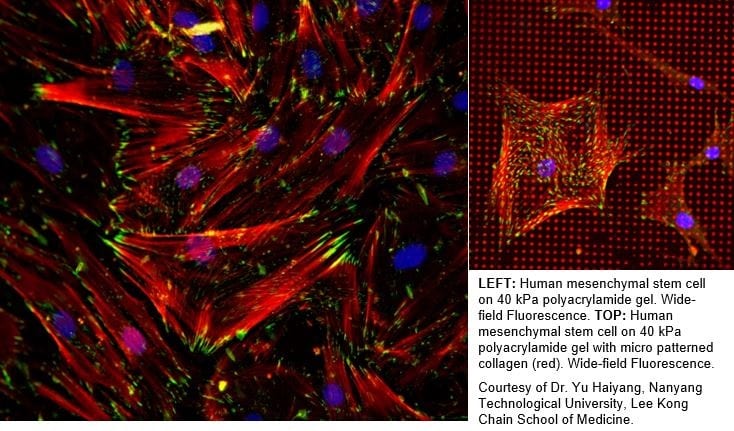
LEFT: Human mesenchymal stem cell on 40 kPa polyacrylamide gel. Wide-field Fluorescence. TOP: Human mesenchymal stem cell on 40 kPa polyacrylamide gel with micro patterned collagen (red). Wide-field Fluorescence.
Courtesy of Dr. Yu Haiyang, Nanyang Technological University, Lee Kong Chain School of Medicine.
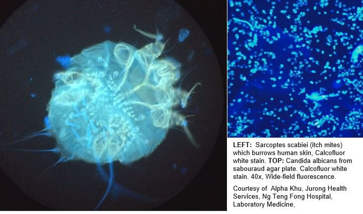
LEFT: Sarcoptes scabiei (itch mites) which burrows human skin, Calcofluor white stain. TOP: Candida albicans from sabouraud agar plate. Calcofluor white stain. 40x, Wide-field fluorescence.
Courtesy of Alpha Khu, Jurong Health Services, Ng Teng Fong Hospital, Laboratory Medicine,
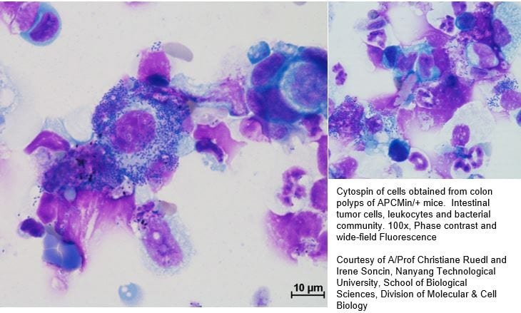
Cytospin of cells obtained from colon polyps of APCMin/+ mice. Intestinal tumor cells, leukocytes and bacterial community. 100x, Phase contrast and wide-field Fluorescence
Courtesy of A/Prof Christiane Ruedl and Irene Soncin, Nanyang Technological University, School of Biological Sciences, Division of Molecular & Cell Biology
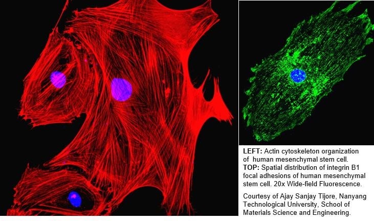
LEFT: Actin cytoskeleton organization of human mesenchymal stem cell. TOP: Spatial distribution of integrin B1 focal adhesions of human mesenchymal stem cell. 20x Wide-field Fluorescence.
Courtesy of Ajay Sanjay Tijore, Nanyang Technological University, School of Materials Science and Engineering.
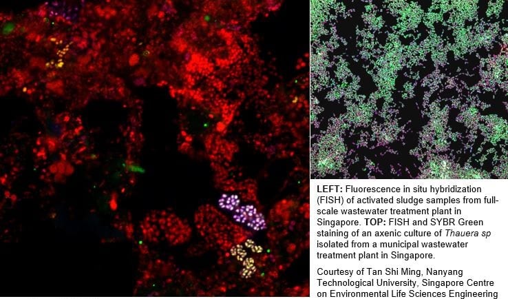
LEFT: Fluorescence in situ hybridization (FISH) of activated sludge samples from full-scale wastewater treatment plant in Singapore. TOP: FISH and SYBR Green staining of an axenic culture of Thauera sp isolated from a municipal wastewater treatment plant in Singapore.
Courtesy of Tan Shi Ming, Nanyang Technological University, Singapore Centre on Environmental Life Sciences Engineering
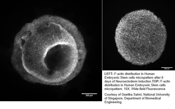
LEFT: F-actin distribution in Human Embryonic Stem cells micropattern after 6 days of Neuroectoderm induction.TOP: F-actin distribution in Human Embryonic Stem cells micropattern. 10X, Wide-field Fluorescence
Courtesy of Geetika Sahni, National University of Singapore, Department of Biomedical Engineering.
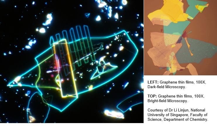
LEFT: Graphene thin films, 100X, Dark-field Microscopy.
TOP: Graphene thin films, 100X, Bright-field Microscopy.
Courtesy of Dr Li Linjun, National University of Singapore, Faculty of Science, Department of Chemistry.
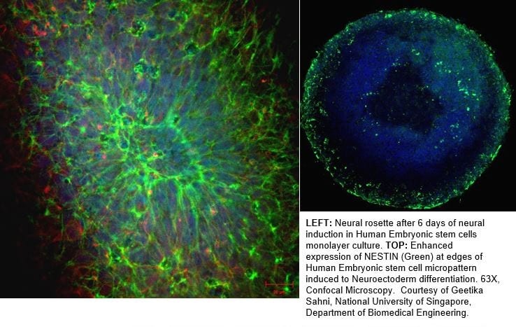
LEFT: Neural rosette after 6 days of neural induction in Human Embryonic stem cells monolayer culture. TOP: Enhanced expression of NESTIN (Green) at edges of Human Embryonic stem cell micropattern induced to Neuroectoderm differentiation. 63X, Confocal Microscopy. Courtesy of Geetika Sahni, National University of Singapore, Department of Biomedical Engineering.
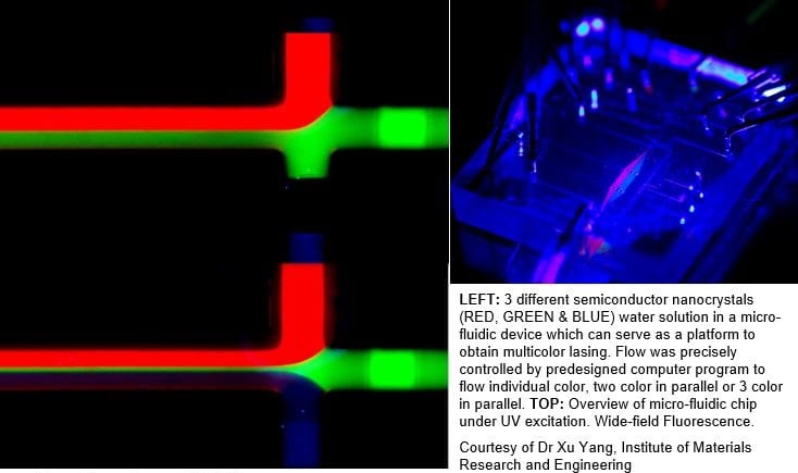
LEFT: 3 different semiconductor nanocrystals (RED, GREEN & BLUE) water solution in a micro-fluidic device which can serve as a platform to obtain multicolor lasing. Flow was precisely controlled by predesigned computer program to flow individual color, two color in parallel or 3 color in parallel. TOP: Overview of micro-fluidic chip under UV excitation. Wide-field Fluorescence.
Courtesy of Dr Xu Yang, Institute of Materials Research and Engineering
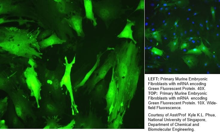
LEFT: Primary Murine Embryonic Fibroblasts with mRNA encoding Green Fluorescent Protein. 40X. TOP: Primary Murine Embryonic Fibroblasts with mRNA encoding Green Fluorescent Protein. 10X. Wide-field Fluorescence.
Courtesy of Asst/Prof Kyle K.L. Phua, National University of Singapore, Department of Chemical and Biomolecular Engineering.
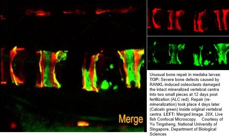
Unusual bone repair in medaka larvae: TOP: Severe bone defects caused by RANKL-induced osteoclasts damaged the intact mineralized vertebral centra into two small pieces at 12 days post fertilization (ALC red). Repair (re-mineralization) took place 4 days later (Calcein green) inside original vertebral centra. LEFT: Merged image. 20X, Live fish Confocal Microscopy. Courtesy of Yu Tingsheng, National University of Singapore, Department of Biological Sciences
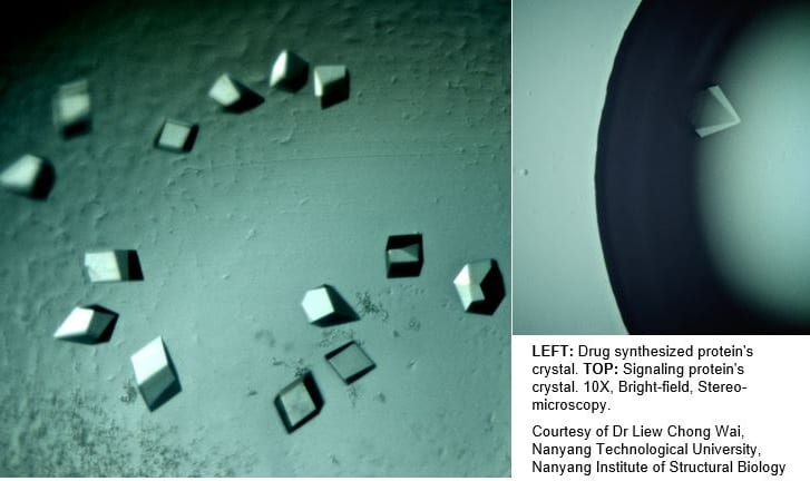
LEFT: Drug synthesized protein’s crystal. TOP: Signaling protein’s crystal. 10X, Bright-field, Stereo-microscopy.
Courtesy of Dr Liew Chong Wai, Nanyang Technological University, Nanyang Institute of Structural Biology