EINST Technology has pioneered its way through the microscopy industry in Singapore. Over the years, we have worked with researchers and academic professional to build a stellar reputation for the company. Beautiful images below are some of the many microscopic images taken by our clients, collaborators and partners which have been proudly incorporated in our yearly EINST calendar.
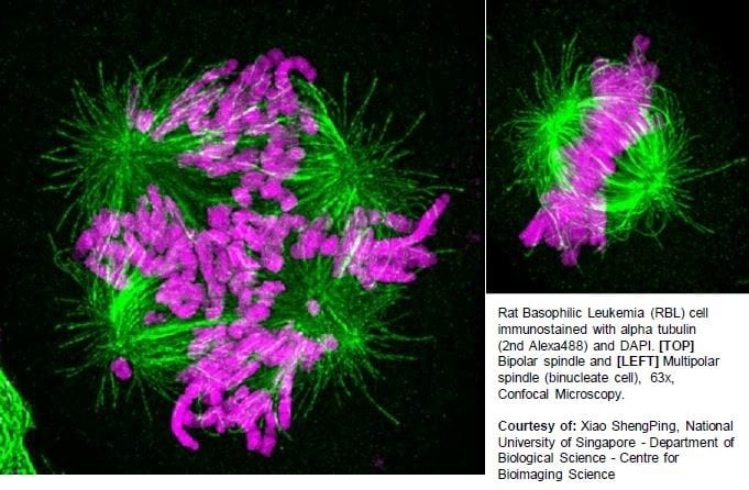
Rat Basophilic Leukemia (RBL) cell immunostained with alpha tubulin (2nd Alexa488) and DAPI. [TOP] Bipolar spindle and [LEFT] Multipolar spindle (binucleate cell), 63x, Confocal Microscopy. Courtesy of: Xiao ShengPing, National University of Singapore – Department of Biological Science – Centre for Bioimaging Science.
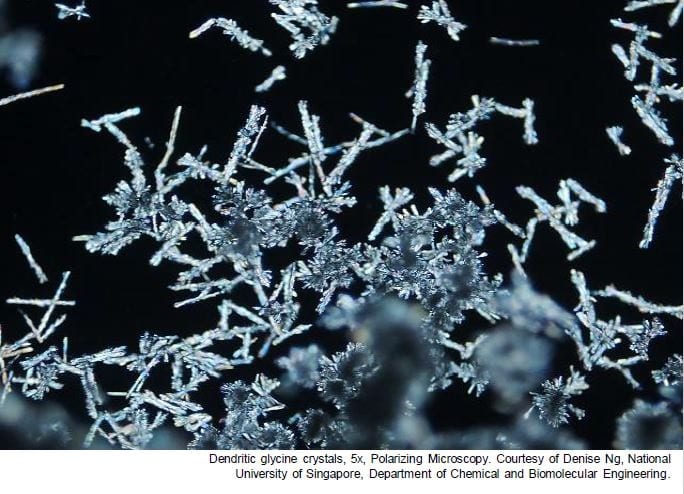
Dendritic glycine crystals, 5x, Polarizing Microscopy. Courtesy of Denise Ng, National University of Singapore, Department of Chemical and Biomolecular Engineering.
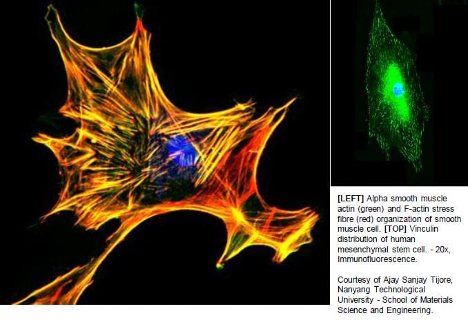
[LEFT] Alpha smooth muscle actin (green) and F-actin stress fibre (red) organization of smooth muscle cell. [TOP] Vinculin distribution of human mesenchymal stem cell. – 20x, Immunofluorescence.
Courtesy of Ajay Sanjay Tijore, Nanyang Technological University – School of Materials Science and Engineering.
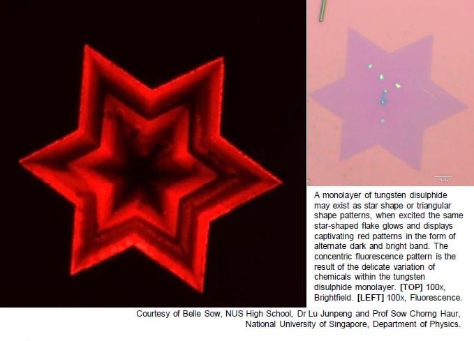
A monolayer of tungsten disulphide may exist as star shape or triangular shape patterns, when excited the same star-shaped flake glows and displays captivating red patterns in the form of alternate dark and bright band. The concentric fluorescence pattern is the result of the delicate variation of chemicals within the tungsten disulphide monolayer. [TOP] 100x, Brightfield. [LEFT] 100x, Fluorescence.
Courtesy of Belle Sow, NUS High School, Dr Lu Junpeng and Prof Sow Chorng Haur, National University of Singapore, Department of Physics.
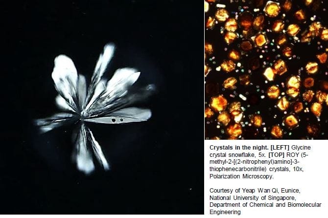
Crystals in the night. [LEFT] Glycine crystal snowflake, 5x. [TOP] ROY (5-methyl-2-[(2-nitrophenyl)amino]-3-thiophenecarbonitrile) crystals, 10x, Polarization Microscopy.
Courtesy of Yeap Wan Qi, Eunice, National University of Singapore, Department of Chemical and Biomolecular Engineering
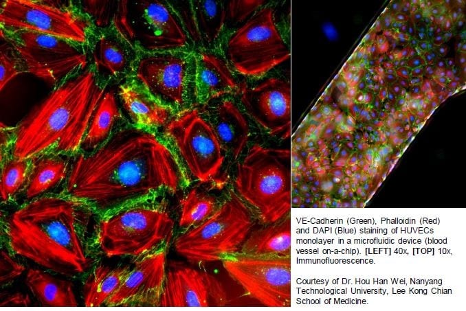
VE-Cadherin (Green), Phalloidin (Red) and DAPI (Blue) staining of HUVECs monolayer in a microfluidic device (blood vessel on-a-chip). [LEFT] 40x, [TOP] 10x, Immunofluorescence.
Courtesy of Dr. Hou Han Wei, Nanyang Technological University, Lee Kong Chian School of Medicine.
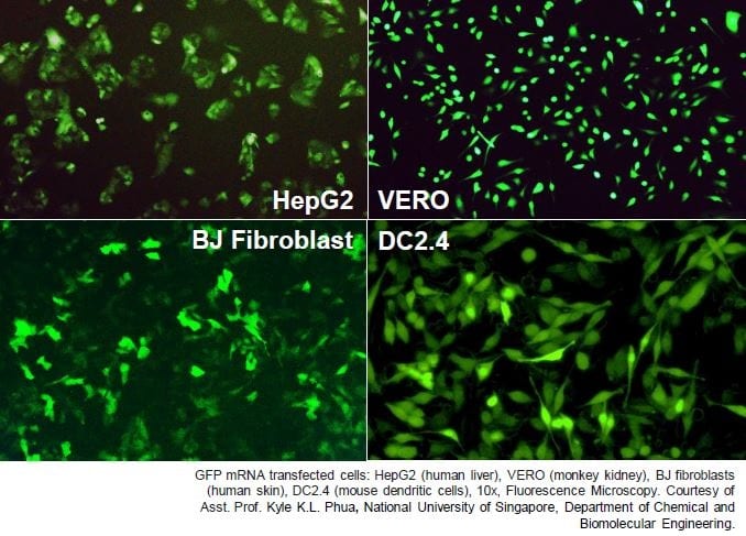
GFP mRNA transfected cells: HepG2 (human liver), VERO (monkey kidney), BJ fibroblasts (human skin), DC2.4 (mouse dendritic cells), 10x, Fluorescence Microscopy.
Courtesy of Asst. Prof. Kyle K.L. Phua, National University of Singapore, Department of Chemical and Biomolecular Engineering
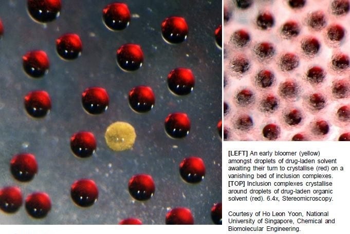
[LEFT] An early bloomer (yellow) amongst droplets of drug-laden solvent awaiting their turn to crystallise (red) on a vanishing bed of inclusion complexes. [TOP] Inclusion complexes crystallise around droplets of drug-laden organic solvent (red). 6.4x, Stereomicroscopy.
Courtesy of Ho Leon Yoon, National University of Singapore, Chemical and Biomolecular Engineering.
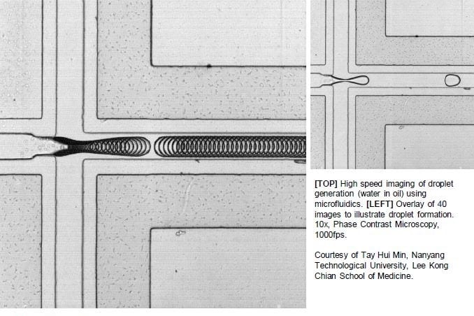
[TOP] High speed imaging of droplet generation (water in oil) using microfluidics. [LEFT] Overlay of 40 images to illustrate droplet formation. 10x, Phase Contrast Microscopy, 1000fps.
Courtesy of Tay Hui Min, Nanyang Technological University, Lee Kong Chian School of Medicine
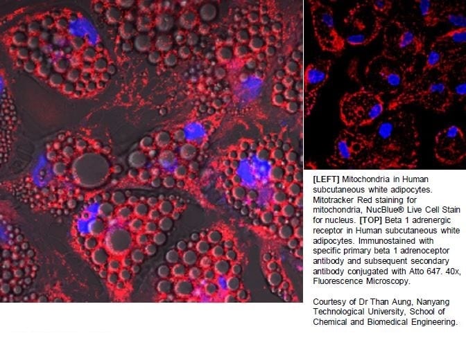
[LEFT] Mitochondria in Human subcutaneous white adipocytes. Mitotracker Red staining for mitochondria, NucBlue® Live Cell Stain for nucleus.
[TOP] Beta 1 adrenergic receptor in Human subcutaneous white adipocytes. Immunostained with specific primary beta 1 adrenoceptor antibody and subsequent secondary antibody conjugated with Atto 647. 40x, Fluorescence Microscopy.
Courtesy of Dr Than Aung, Nanyang Technological University, School of Chemical and Biomedical Engineering
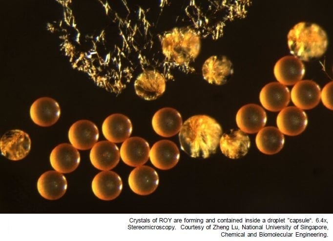
Crystals of ROY are forming and contained inside a droplet “capsule“. 6.4x, Stereomicroscopy. Courtesy of Zheng Lu, National University of Singapore, Department of Chemical and Biomolecular Engineering.
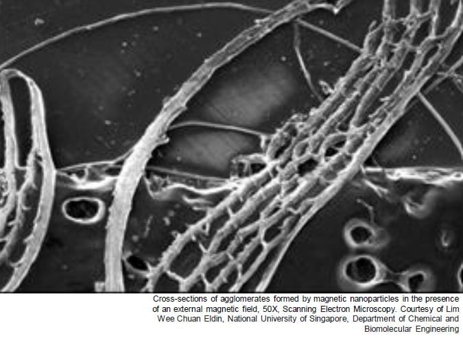
Cross-sections of agglomerates formed by magnetic nanoparticles in the presence of an external magnetic field, 50X, Scanning Electron Microscopy. Courtesy of Lim Wee Chuan Eldin, National University of Singapore, Department of Chemical and Biomolecular Engineering