Enquiry Form
Enquiry Form

TOMOCUBE - Holographic Microscope

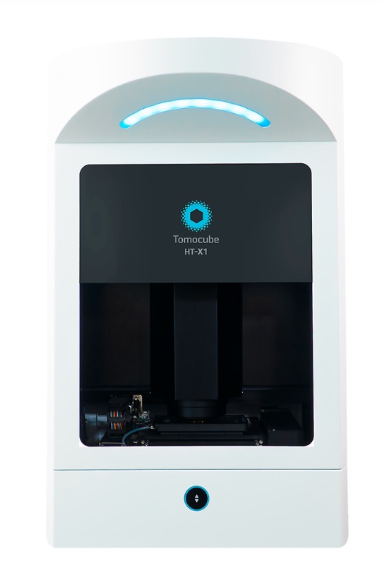
Tomocube HT-X1 Holotomography

Tomocube HT-X1 Holotomography

By capturing the intrinsic refractive index (RI) of cells using a low level of light intensity, Holotomography has emerged as a unique solution for live cell imaging that surpasses the compromise between obtaining high image quality and maintaining healthy cells.

Empowering your exploration. Redefining the limits.


The lastest generation of Holotomography – Tomocube HT-X1 – utilizes a low-coherence light source. This groundbreaking advancement unlocks label-free 3D and 4D quantitative live cell imaging on diverse multi-well plates with improved resolution and free of laser-induced speckle noise.

The new system works well with a wide range of biological samples, including confluent cell sheets and thick specimens. It provides not only high-content 3D visualization but also quantitative data for individual cells and their intracellular structures, such as the volume, surface area, and dry mass.


Multiplying Knowledge from 2D to 4D and Beyond

Observations of cellular morphology and activity are typically based on labeling of target molecules. These methods are invasive, affect the nature of the target molecules and potentially interfere with their biological relevance. In addition, these forms of label are not effective in providing quantitative information. The tools used to visualize labels are generally damaging to cells as often laser-based illumination is used to excite fluorochromes which in turn causes phototoxic damage to the cells. The “holy grail” of imaging is to visualize cells without labeling to ensure the cells behave and grow normally. Holotomography (HT) does this by capturing the intrinsic light scattering properties of cellular materials using very low levels of light intensity, just enough to allow the light to pass through the cell. In doing so, the refractive index (RI) information of structures within the cell can be collected and selectively pseudo-colored to reveal the cell and its organelles. In capturing the RI distribution, the Tomocube HT-X1 Holotomography can also provide quantitative data in 3D such as the volume, surface area, and dry mass of cells and their intracellular structures.
 

Correlative HT to FL

 
[video width="390" height="400" mp4="https://www.einstinc.com/wp-content/uploads/2023/08/holomotography-and-fluorescence.mp4"] [/video]

Holotomography and Fluorescence

[video width="390" height="400" mp4="https://www.einstinc.com/wp-content/uploads/2023/08/Flourescence.mp4"][/video]

Fluorescence

View More
Holotomographic microscopy HT 2

Holotomographic microscopy HT 2

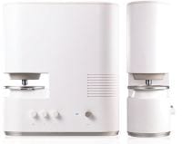
The HT-2 series opens a new era of 3D correlative imaging, combining the holotomography and fluorescence methods. The HT-2 allows the viewing of a 3D RI tomographic image of a living cell with minimal damage, while at the same time targeting consumer interest by allowing for fluorescence labeling in the vary same cell.  

HT-2 : Holotomographic microscopy with 3D fluorescence imaging capability

 
Research and diagnosis in the life sciences depends on the information that can be found using cellular analysis. However, current microscopy techniques limit the quantity and quality of information available to researchers and clinicians The HT-2, a microscope that displays both 3D holograms and 3D fluorescence imaging, is able to overcome these limitations through use of new and existing technology. Its abilities can enable researchers and clinicians to break into new frontiers of understanding, diagnosing, and treating disease. HT-2 is the world’s first microscopy combining both holotomography and 3D fluorescence imaging in one unit. The HT-2 is an update to our HT-1 microscope. The HT-1 microscope uses holotomographic technology to measure the 3D and 4D refractive indexes using a dynamic micromirror device (DMD) to add unprecedented stability and precision to the performance of the microscope. These innovations allow for quantitative, label free imaging of cells. The HT-2 has all the capabilities of the HT-1, but combines the holotomographic imaging capabilities of the original version with the added ability to perform fluorescence microscopy.
View More
Holotomographic Microscopy – HT 1

Holotomographic Microscopy – HT 1

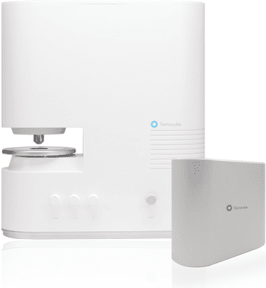
HT-1 is a new microscope designed to let researchers view live cell using holotomograhpy technology. One of the world's first microscopes of its kind, the HT-1 delivers non-invasive, real-time quantitative data with the highest resolution on the market today. Holotomographic Microscopy - HT 1

Revolutionary holotomographic (3D holographic) microscopy opens new era for label-free live cell imaging

Cellular analysis plays a crucial role in a wide variety of research and diagnostic activities in the life science. However, the information available to researchers and clinicians is limited by current microscopy techniques. An innovative new tool – Holotomographic microscopy – can overcome many of these limitations and open new vistas for researchers and clinicians to understand, diagnose and treat human diseases. Holotomographic Microscopy – New era of microscopy Tomocube’s holotomography series utilize optical diffraction tomography (ODT), which enables users to quantitatively and noninvasively investigate biological cells and thin tissues. ODT reconstructs the 3D refractive index (RI) distributions of live cells and by doing so, provides structural and chemical information about the cell including dry mass, morphology, and dynamics of the cellular membrane.

TomoStudio™, Software platform for 2D/3D/4D image analysis

TomoStudio™, HT-1 operating SW, controls the system and visualizes the captured image in various ways. This flexible user interface provides fast imaging capability and 2D/3D/4D visualization of the cellular image based on 3D RI distributions of the cells and tissues. follow this link. TomoStudio™

Smart technology for holotomography

HT-1 use holotomography technology which measures the 3D and 4D refractive index (RI) tomograms. This new technology enables quantitative, label-free measurements of live, unstained cells and tissue samples. With the dynamic micromirror device (DMD), we have eliminated moving parts from light path while image-capturing and has added unprecedented stability and precision on the outstanding performance of our product. If you want to get more information about our technology, follow this link. Technology
View More