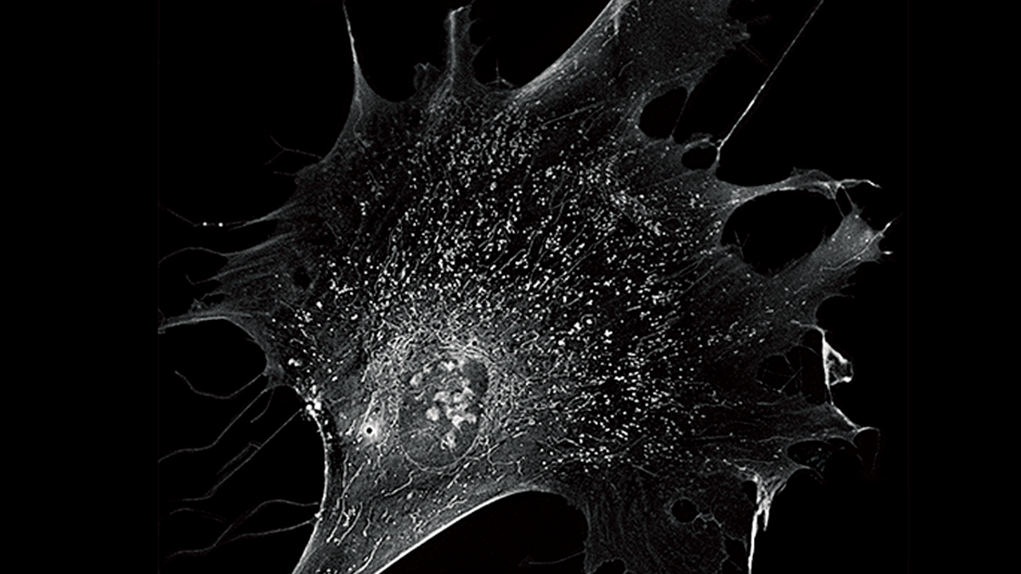
By capturing the intrinsic refractive index (RI) of cells using a low level of light intensity, Holotomography has emerged as a unique solution for live cell imaging that surpasses the compromise between obtaining high image quality and maintaining healthy cells.
Observations of cellular morphology and activity are typically based on labeling of target molecules. These methods are invasive, affect the nature of the target molecules and potentially interfere with their biological relevance. In addition, these forms of label are not effective in providing quantitative information. The tools used to visualize labels are generally damaging to cells as often laser-based illumination is used to excite fluorochromes which in turn causes phototoxic damage to the cells.
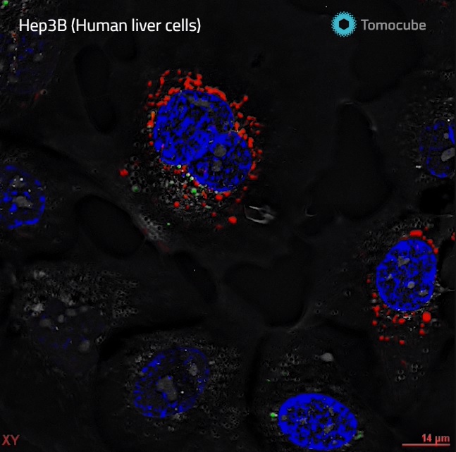
The “holy grail” of imaging is to visualize cells without labeling to ensure the cells behave and grow normally. Holotomography (HT) does this by capturing the intrinsic light scattering properties of cellular materials using very low levels of light intensity, just enough to allow the light to pass through the cell. In doing so, the refractive index (RI) information of structures within the cell can be collected and selectively pseudo-colored to reveal the cell and its organelles.
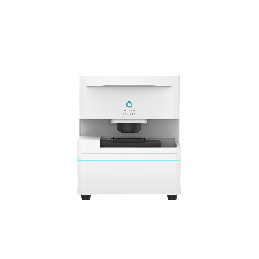
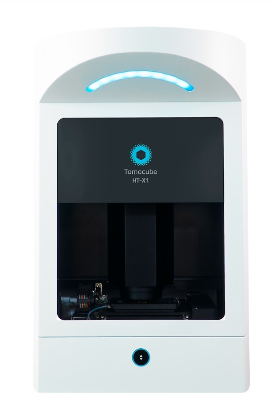
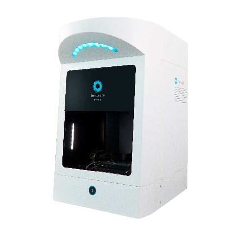
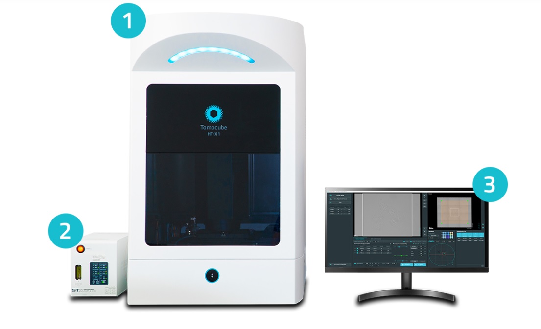
In capturing the RI distribution, the Tomocube HT-X1 Holotomography can also provide quantitative data in 3D such as the volume, surface area, and dry mass of cells and their intracellular structures.
Holotomography and Fluorescence |
Fluorescence |
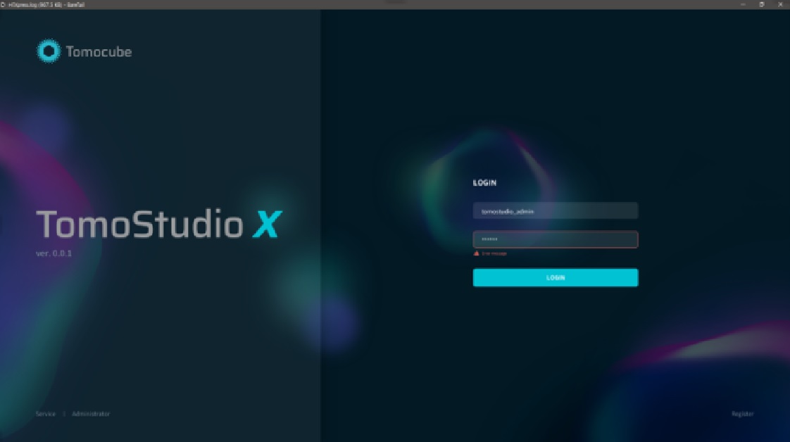
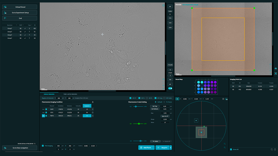
As the control software for the HT-X1, TomoStudio X is designed to operate the system as you design the experiment. TomoStudio X offers flexible workflows for multi-dimensional image acquisition of holotomography and fluorescence in multi-well imaging conditions. All the essential control and status of the system are displayed all at one glance.
TomoStudio X works in concert with the HT-X1 platform to visualize and analyze RI tomograms. This intuitive, easy-to-use software empowers users with complete system control, allowing them to handle even the most complex experiments through simple mouse clicks. Researchers can easily set up complex time-lapse sequences, perform large-area tile imaging and stitching tasks, and image multiple points with ease using this software.