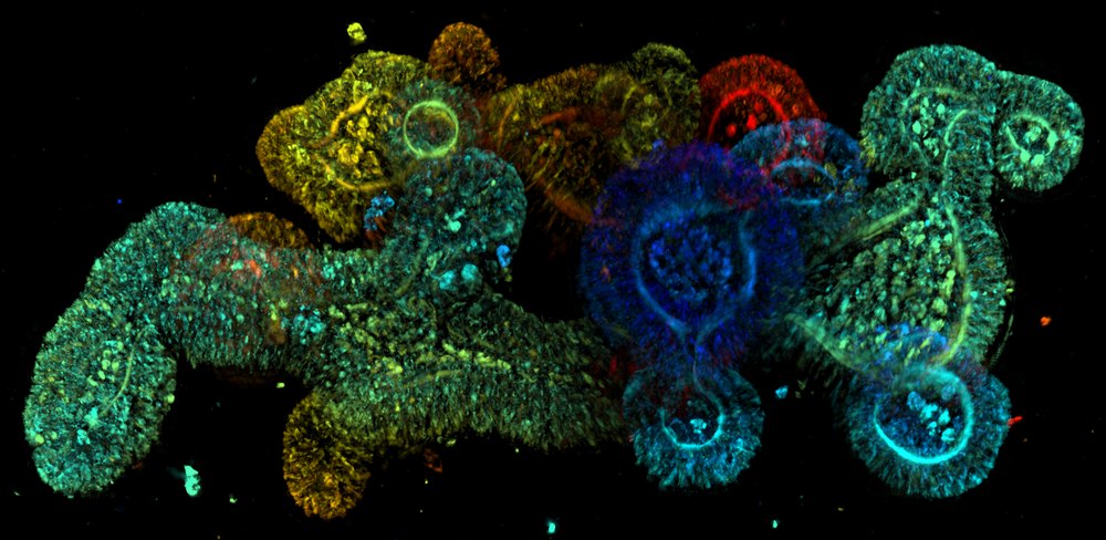
Label-free 3D high-content imaging for organoids
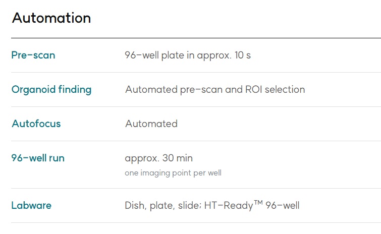
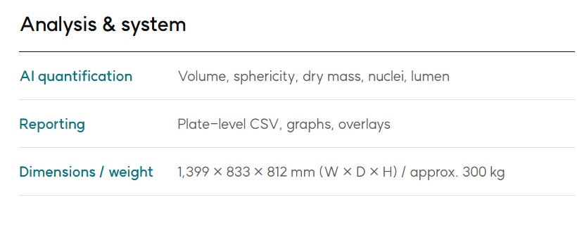
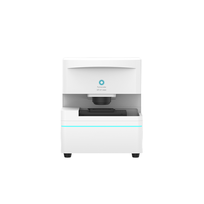
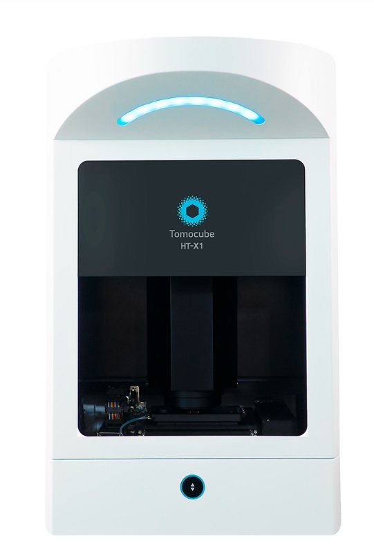
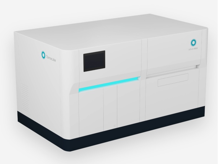
HT-X1 MAX is an imaging system for organoids, based on holotomography (HT). It images whole organoids in 3D without staining, keeps them in culture for time-lapse, and turns each image into quantitative readouts, in formats from dishes up to 96-well plates.
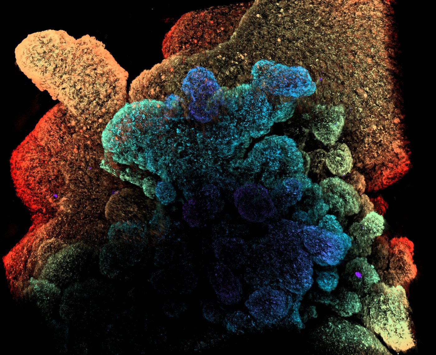
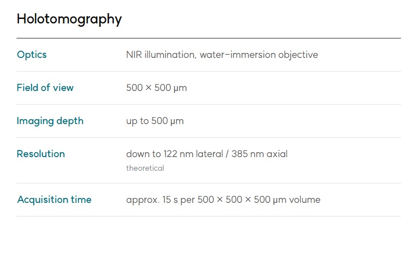
HT images through organoids up to 500 μm. Analysis is not limited to smaller organoids.
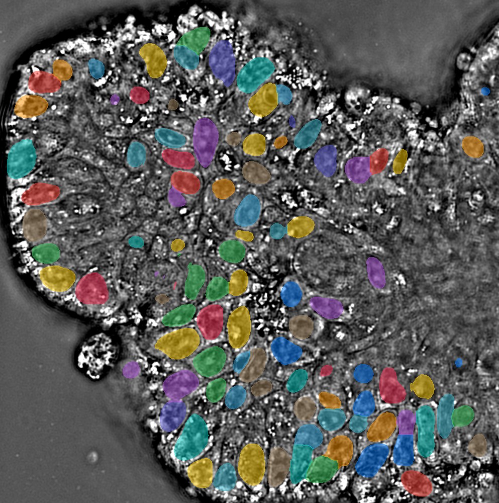
HT resolves organoid structure, nuclei, the cell layer and the lumen without labels. Your fluorescence channels stay free for the markers you choose.
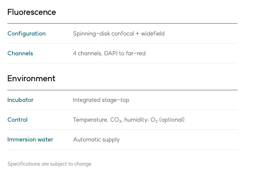
The stage-top incubator holds temperature, CO₂ and humidity. Organoids stay in culture throughout imaging, for hours to days.
Endpoint assays use up a separate set of organoids at every time point. Imaging the same organoids follows the whole response with far fewer samples. Each organoid gets its own trajectory, not a population average.