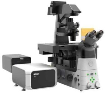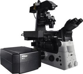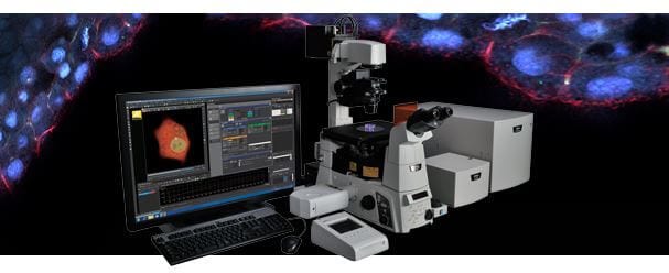
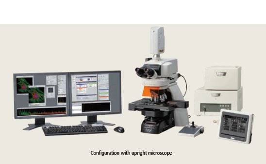
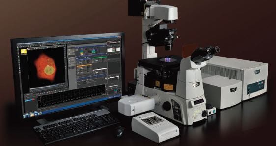
Simple and Robust. The C2+ is designed as an essential microscopy tool for the laboratory, providing powerful and robust imaging capabilities….
Point scanning confocal microscope system offering powerful new features and vastly expanded spectral imaging capabilities.
Built on a reputation of incredible stability coupled with superior optical technologies, the C2+ with its host of functions and various analytical capabilities is the perfect tool for a new microscope, or as a new accessory to a Nikon imaging system. Now fully controlled by NIS-Elements imaging software, the system includes four channel confocal fluorescence imaging, and vastly expanded spectral capabilities with the ability to capture and unmix data acquired at any channel resolution across the entire detector bandwidth.
The C2+ fits all Nikon microscopes with the smallest scan head footprint on the market. The system employs high precision mirrors and optically ideal circular pinholes, enabling noiseless, high contrast and high quality confocal imaging. The spectral detector of the C2+ enables high speed imaging using simultaneous quantitative 32-channel acquisition. Signal loss has been minimized while imaging of fluorescence spectra in real colors is possible by host of innovations for accurately correcting spectral data.


CFI Apo 40xWI λS, NA1.25 (left)
CFI Apo LWD 40xWI λS, NA1.15 (middle)
CFI Apo 60x Oil λS, NA1.4 (right)
These high NA objectives are ideal for confocal imaging with correction of chromatic aberrations over a wide wavelength range, from ultra violet to infrared. Transmission is increased through the use of Nikon’s exclusive Nano Crystal Coat technology.
Acquisition of a 32-channel spectral image (512 x 512 pixels) with a single scan in 0.6 second is possible. Moreover, 512 x 32-pixel images can be captured at 24 fps.
Accurate, High-speed Unmixing
Accurate spectral unmixing provides maximum performance in the separation of closely overlapping fluorescence spectra and the elimination of autofluorescence. Superior algorithms and high-speed data processing enable real time unmixing during image acquisition.


The C2+ handles simultaneous 3-channel fluorescence or simultaneous 3-channel and diascopic DIC observation. High quality DIC images and fluorescence images can be superimposed to aid in image analysis such as locating fluorescence labels.
Enhanced Spectral Imaging
Acquisition of a 32-channel spectral image (512 x 512 pixels) with a single scan in 0.6 second is possible. Moreover, 512 x 32-pixel images can be captured at 24 fps.
Accurate, High-speed Unmixing
Accurate spectral unmixing provides maximum performance in the separation of closely overlapping fluorescence spectra and the elimination of autofluorescence. Superior algorithms and high-speed data processing enable real time unmixing during image acquisition.

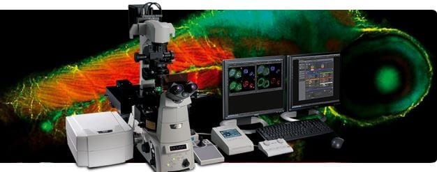
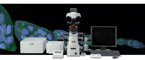
NIS-Elements C control software enables integrated control of the confocal imaging system, microscope and peripheral devices with a simple and intuitive interface. Diverse reliable analysis functions are also available.
| Compatible Laser: | Solid-state laser: 405 nm, 440 (445) nm, 488 nm, 561 (594) nm, 638 (640) nmAr laser (457 nm/488 nm/514 nm), HeNe laser (543 nm)Solid-state laser: 405 nm, 440 (445) nm, 488 nm, 561 (594) nm, 638 (640) nmAr laser (457 nm/488 nm/514 nm), HeNe laser (543 nm) |
|---|---|
| Laser Unit: | C-LU3EX 3-laser unit EX (AOM or manual modulation), LU4A 4-laser unit (AOTF modulation) |
| Standard Detector: | Wavelength: 400-750nm Detector: 3 PMT Filter cube: 2 filter cubes |
| Diascopic Detector (option): | Wavelength: 400-700nm Detector: 1 PMT |
| Spectral Detector (option): | Number of channels: 32 Wavelength: 400-750nm Wavelength resolution: 2.5 nm/5 nm/10 nm, wavelength range variable in 0.25nm steps Unmixing: High-speed unmixing, precision unmixing |
| Scanning: | Scanning range: square inscribed in a ø18mm circle
With 3 channel fluorescent detector: With spectral detector: |
| Scanning Mode: | X-Y, X-Z, XY rotation, zoom, ROI, XYZ, time lapse, stimulation, multipoint, image stitching (large image) |
| Pinhole: | Circular shape, 6 size |
| Image Bit Depth: | 12 bits |
| Compatible Microscopes: | ECLIPSE Ti-E/Ti-U inverted microscope ECLIPSE Ni-E (focusing nosepiece type/stage focusing type) Ni-U upright microscope ECLIPSE FN1 fixed stage microscope AZ100 multi-purpose zoom microscope |
| Z Step: | Ti-E: 0.025 μm, FN1 stepping motor: 0.05 μm, Ni-E: 0.025 μm |
| NIS-Elements C Software: | Acquisition/Visualization/Image Processing/Analysis: 3D volume/orthogonal viewing and rendering, large image stitching, multi-point timelapse, spectral unmixing, DAQ and I|O control, timelapse analysis, deconvolution, ratio analysis, morphology segmentation, AVI and MOV file outputApplications: FRAP, FLIP, FRET, photo activation, colocalization, three-dimensional time-lapse imaging, multipoint time-lapse imaging |
*1 Fast mode is compatible with zoom 8-1000x. It is not compatible with Rotation, Crop, ROI, Spectral Imaging and Stimulation.
 Bioengineering
 Biomechanics
 Cardiovascular Research
 Developmental Biology/Embryology
 Neurobiology / Neuroscience
 Osteology
 Vascular Research