Improving on Perfection
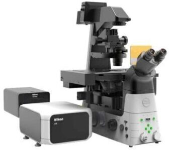
Confocal microscopes have been commercially available now for over 25 years. How can newer iterations of a fundamentally simple instrument continue to innovate? What changes can redefine how a confocal is used, and what data can be collected? Introducing the Nikon AX/AX R Confocal Microscope System, our 10th generation point scanning confocal, giving you more of everything: Leveraging Artificial Intelligence (AI), expanding the number of colors, improving pixel density, sensitivity and speed.
These are significant additions in terms of expanding the range of experiments possible with a point scanning confocal, while increasing the usability and functionality of the instrument, all in a modular and upgradable platform.
Nikon AX is the new standard in confocal imaging.
The newly developed Nikon Spatial Array Confocal (NSPARC) detector utilizes an ultra-low noise detector array to collect a two-dimensional image at each scanned point. This method of image scanning microscopy (ISM) improves signal-to-noise ratio by increasing the available signal level while simultaneously allowing imaging with lower excitation power.
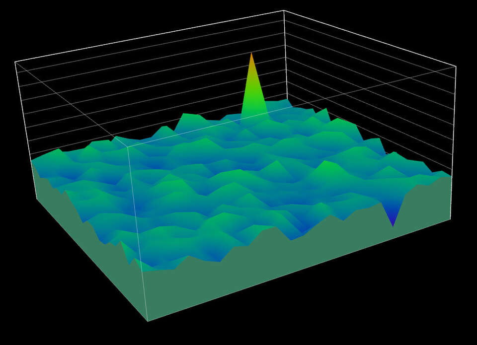
Single-photon sensitivity and array detection extend the capabilities of the AX system by revealing unseen details in every image, while array detection pushes the boundaries of resolution beyond the theoretical limits.
| AX | AX R | |
|---|---|---|
| Scan Head | 25 mm FOV galvano scanner Up to 8192 x 8192 pixels Up to 10 fps at 512 x 512 pixels Pixel dwell time up to 0.2 microseconds Supports bidirectional imaging and line-scan imaging |
25 mm FOV resonant scanner 25 mm FOV galvano scanner Up to 2048 x 2048 pixels for 2K (1024 x 1024 pixels for 1K) Up to 720 fps at 2048 x 16 pixels for 2K (720 fps at 1024 x 16 pixels for 1K) 30 fps at 2048 x 512 pixels for 2K (1024 x 512 pixels for 1K) Supports bidirectional imaging and line-scan imaging |
| Scan Head input/output port | One FC fiber laser input port and one FC fiber signal output port An additional FC fiber laser input port and FC fiber signal output port can be optionally added |
|
| Laser | Up to 8 visible lasers Compatible range: 405-750 nm |
|
| Dichroic Mirror | Up to 6 customizable mirrors | |
| DUX-VB Detector | 2 or 4 channels Freely tunable emission bands with ± 1nm accuracy and up to 66 discrete spectral channels Up to 12 bandpass filters Multi-alkali PMT or GaAsP PMT options |
|
| DUX-ST Detector | 2 or 4 channels Up to 18 bandpass filters Multi-alkali PMT or GaAsP PMT options |
|
| NSPARC Detector Unit | Lateral resolution 100 nm*1, Axial resolution 300 nm*1 Equipped with SPPC (Single Pixel Photon Counter) array detector Up to 7 barrier filters can be mounted (supports excitation at 405 nm, 445 nm, 488 nm, 514 nm, 561 nm, 594 nm and 640 nm) With galvano scanner: Can be used with X resolution of 64 to 8192 pixels, Y resolution of 2 to 8192 pixels With resonant scanner: Can be used with X resolution of 256, 512 and 1024 pixels, Y resolution of 128 to 1024 pixels*2 |
|
| Diascopic Detector | Compact PMT detector | |
| Pinhole | Continuously variable | |
| FOV | Maximum 25 mm diameter (circle) inscribed by a rectangle | |
| Z step | Ti2-E: 0.01 μm, 0.02 μm (with encoder control), FN1 stepping motor: 0.05 μm, Ni-E: 0.025 μm | |
| Compatible microscopes | Ti2-E inverted microscope with maximum FOV of 25 mm Ni-E and FN1 upright microscopes with maximum FOV of 25 mm |
|
| Options | Photostimulation (point raster or digital micromirror) Fluorescence lifetime imaging including fast FLIM Piezoelectric Z (or XYZ) Environmental chamber or enclosure Other modalities such as TIRF, N-STORM or N-SIM S |
|
| Software | Nikon NIS-Elements C Optional modules available Up to 16 bit images (65,536 gray levels with fine integration) with multiple file output options |
|
| Control Workstation | Microsoft Windows® 10 64bit Professional with GPU-accelerated graphics card | |
| Recommended installation conditions | Temperature 23 ± 5˚C, Humidity 70% RH or less (no condensation) | |
*1 These values were measured using 40 nm diameter beads for lateral resolution and 100 nm diameter beads for axial resolution with excitation by a 488 nm laser. Actual resolution depends on laser wavelength and optical configuration.
*2 Resolution of 2048 pixels cannot be set.
Key Features
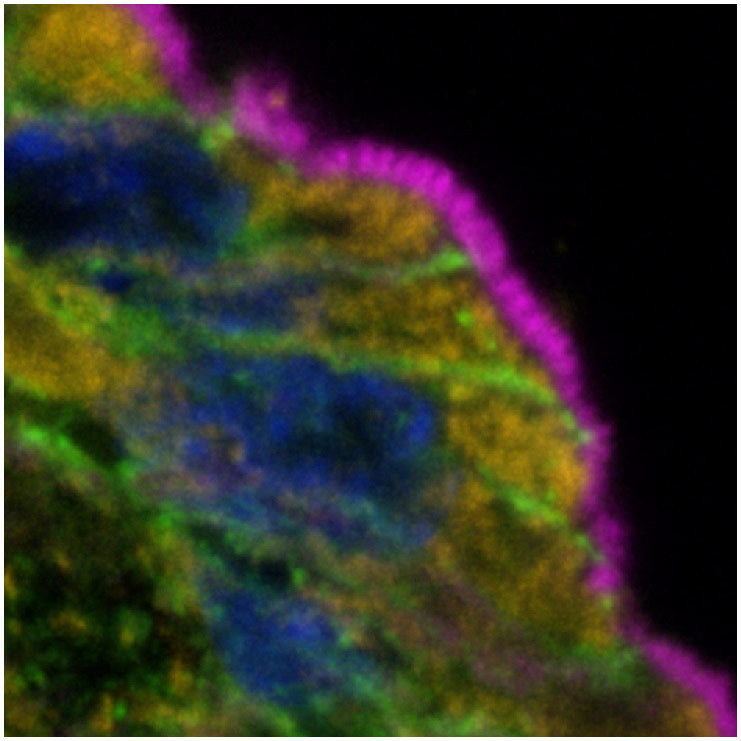
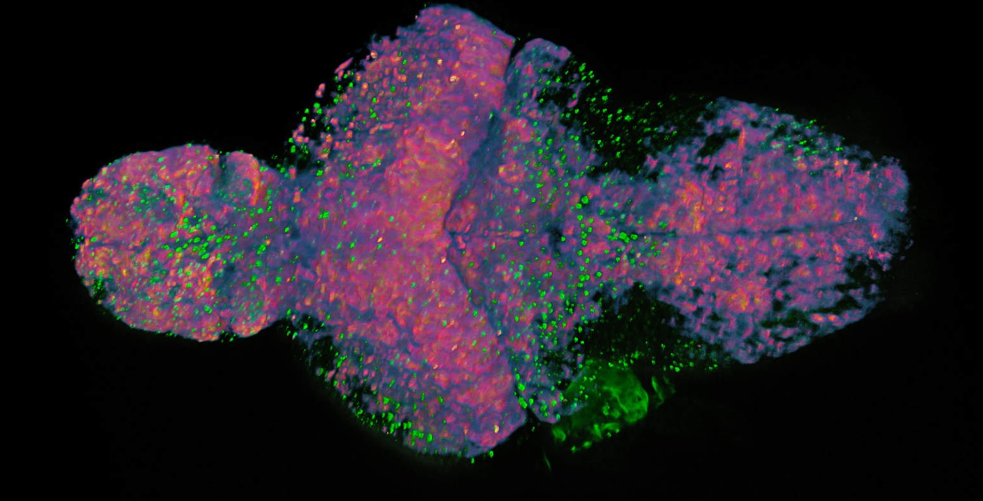
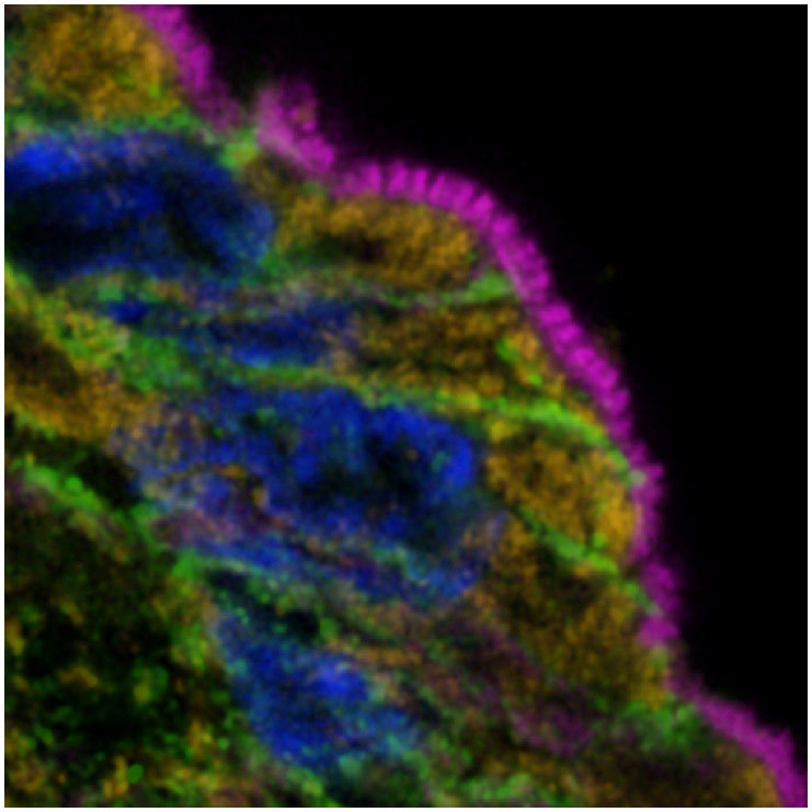
3D Confocal Imaging with Super Resolution
NSPARC’s array detection provides spatial sampling of the confocal point spread function in every pixel. This method detects information that can be utilized to extract enhanced resolution performance both laterally and axially. Built-in variable emission optics increase flexibility to allow users to replicate the function of a pinhole. This means the ability to prioritize enhanced optical sectioning or increased overall signal collection depending upon specific experimental needs.
The variable emission optics allow Nikon to offer many compatible low and high mag objectives to match specimens. This includes silicon immersion objectives which minimize refractive index mismatches, improving overall system performance.
Coupled with the AX/AX R’s ultra-large 25mm FOV, the system supports a wide selection of objectives capable of obtaining image data from large overviews down to extremely fine details, which can then be measured and analyzed.
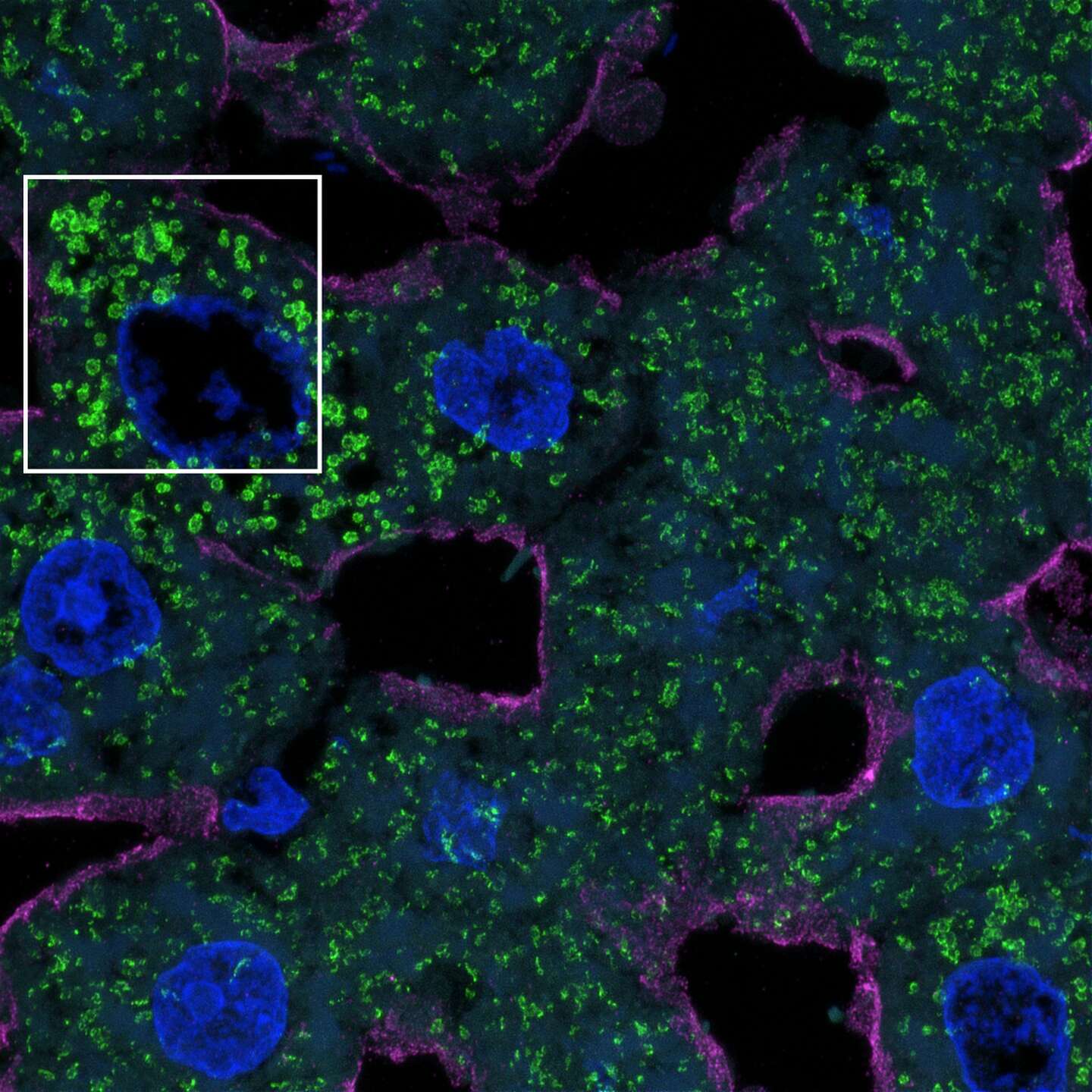
A New Level of Low Noise, High Sensitivity Detection
An extremely low noise profile along with incredible sensitivity renders outstanding results even under demanding imaging conditions, including high speed resonant imaging.
As an added benefit, additional spatial information is acquired for every pixel using the detector array, which can be used to further enhance resolution and brightness.
NSPARC Noise Profile |
GaAsP Noise Profile
|
Comparing NSPARC and GaAsP bias images (acquisition with no light to measure noise) reveal extremely low standard deviation in pixel-to-pixel intensity, significantly lower than GaAsP or PMT detectors.
| NSPARC
|
Traditional Detector
|
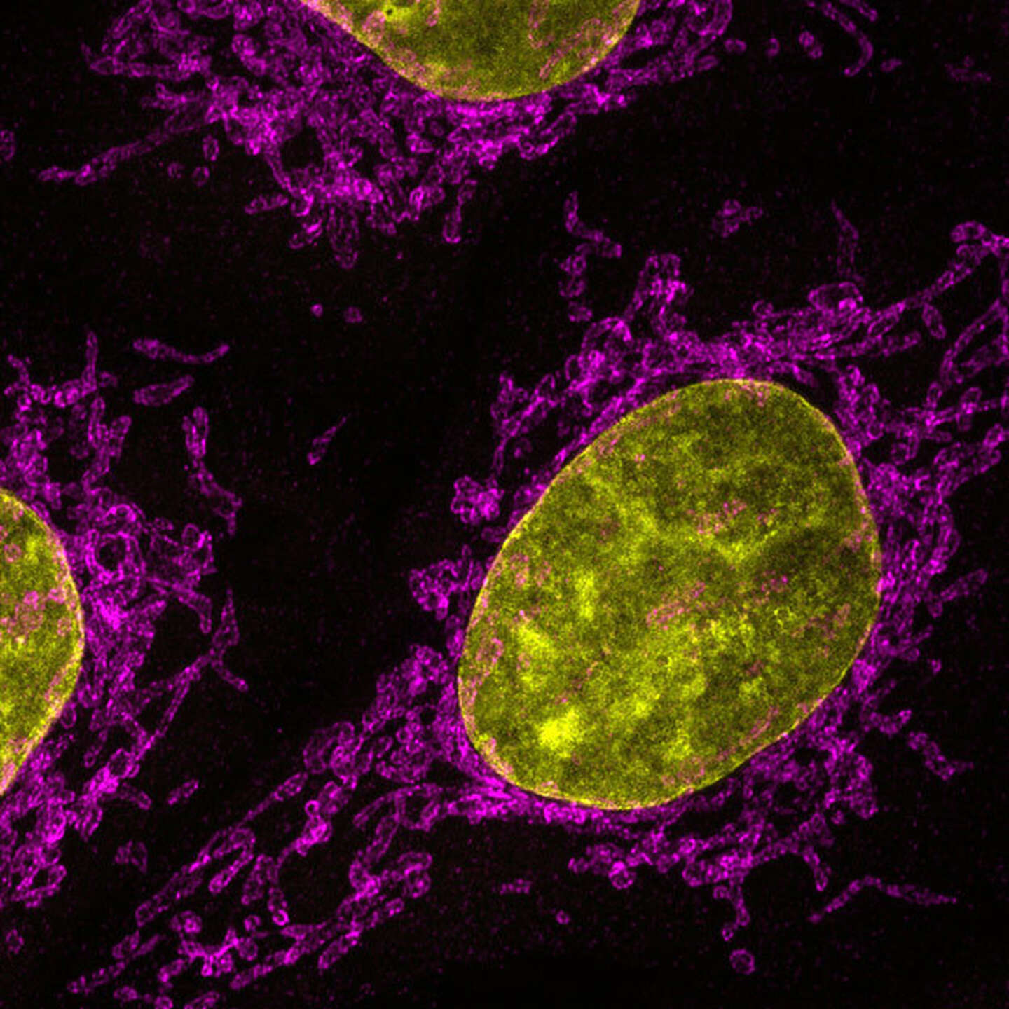
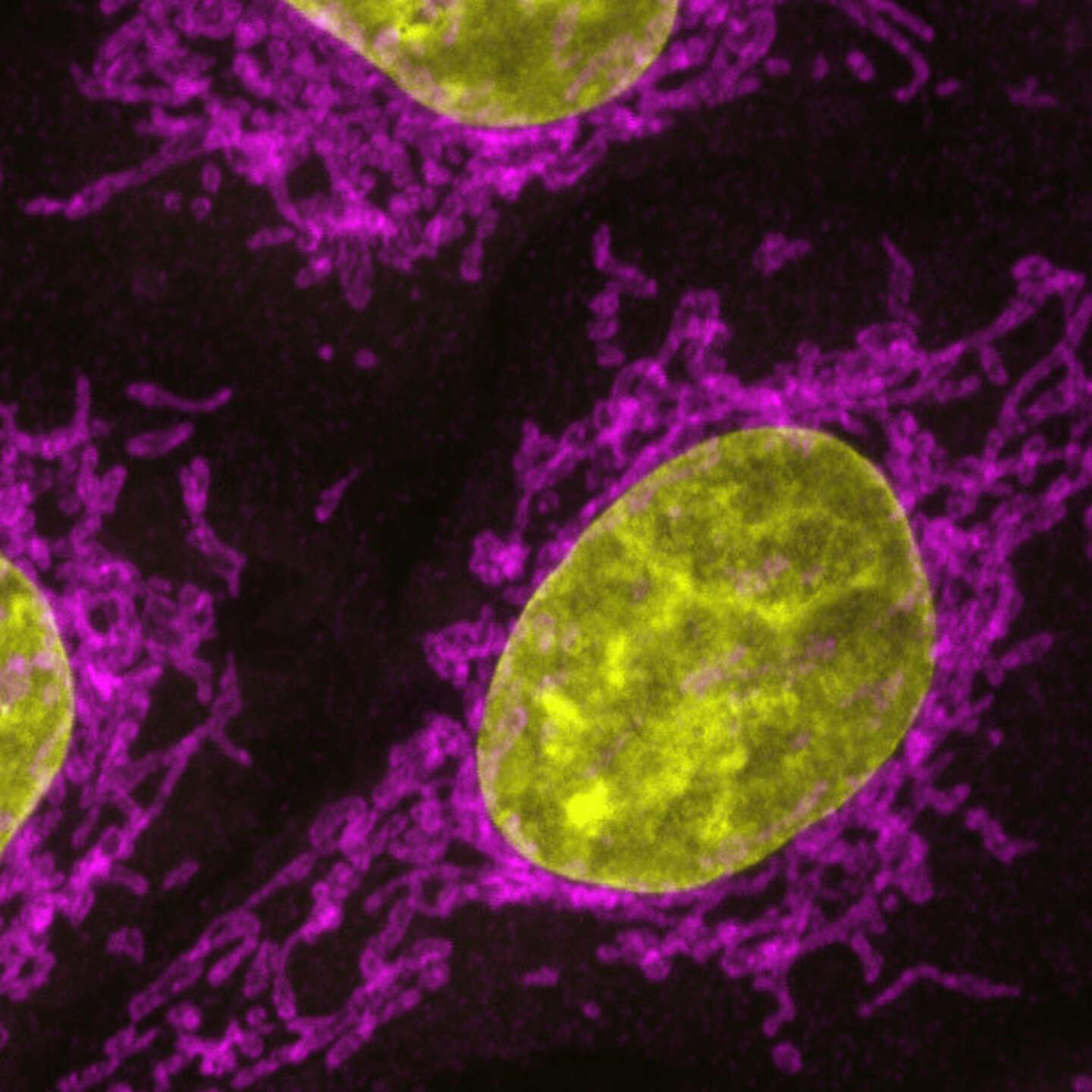
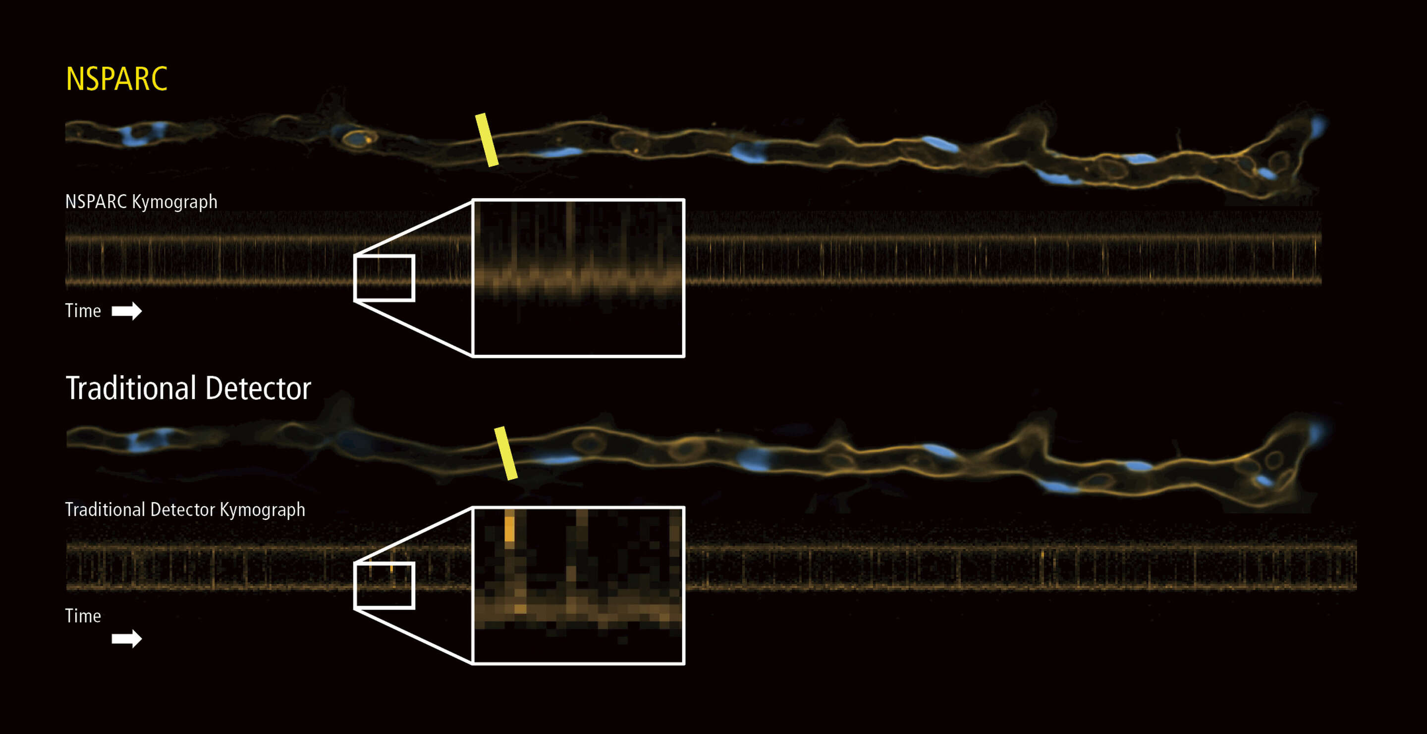
The extremely low noise of NSPARC detectors and the additional spatial data collection per pixel result in high signal-to-noise, sharper images compared to standard GaAsP or PMT images.
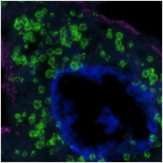
NSPARC Spatial Array Detector Technology
Where a point scanning detector traditionally produces an intensity output only for each pixel, NSPARC comprises an array of 25 detectors that operates more like an extremely sensitive camera: two-dimensional spatial information from each scanned pixel is collected by the detector.
Optical lenses direct emission light to the detector, allowing it to be used with various objectives and magnifications, while simultaneously allowing the user to define the size of the illumination spot on the detector array. This enables oversampling of the conventional single airy unit emission from the confocal plane.
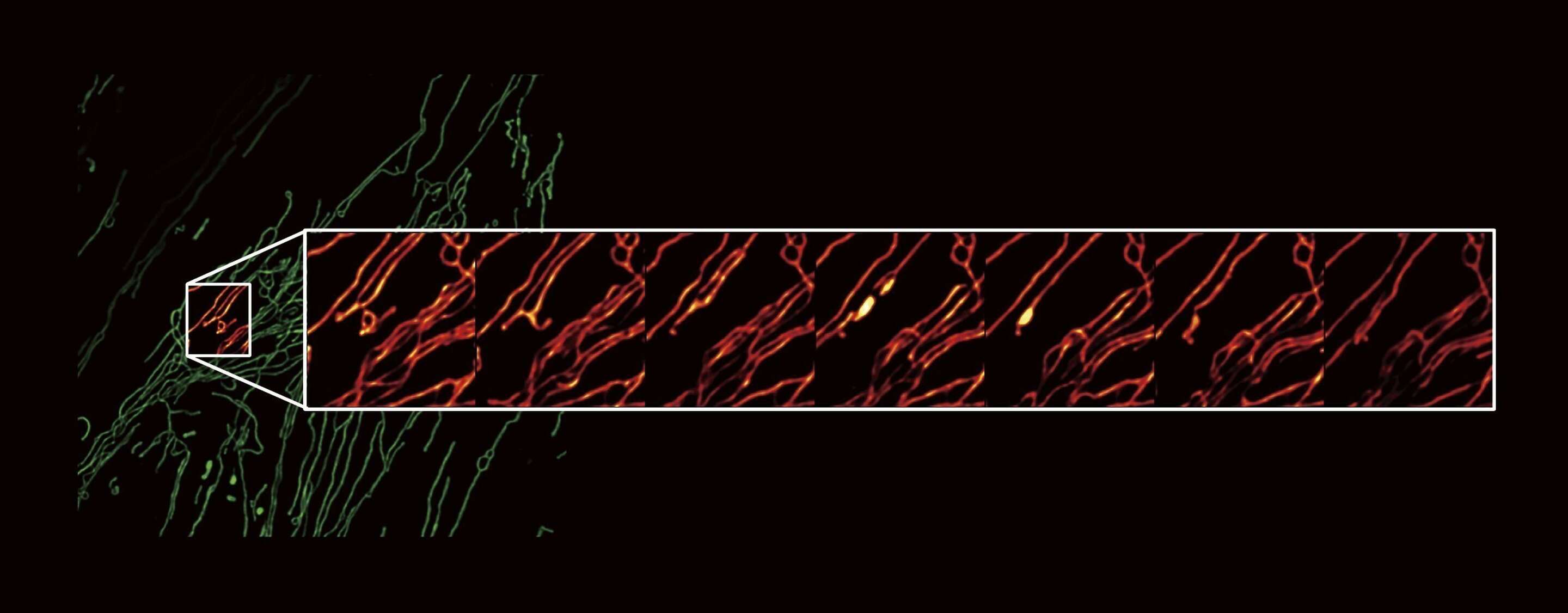
This two-dimensional information is immediately used to obtain ultra-fine structural information, which is lost in conventional detection.
Each pixel from NSPARC contains spatial information, which can be used to reconstruct fine details in a result image.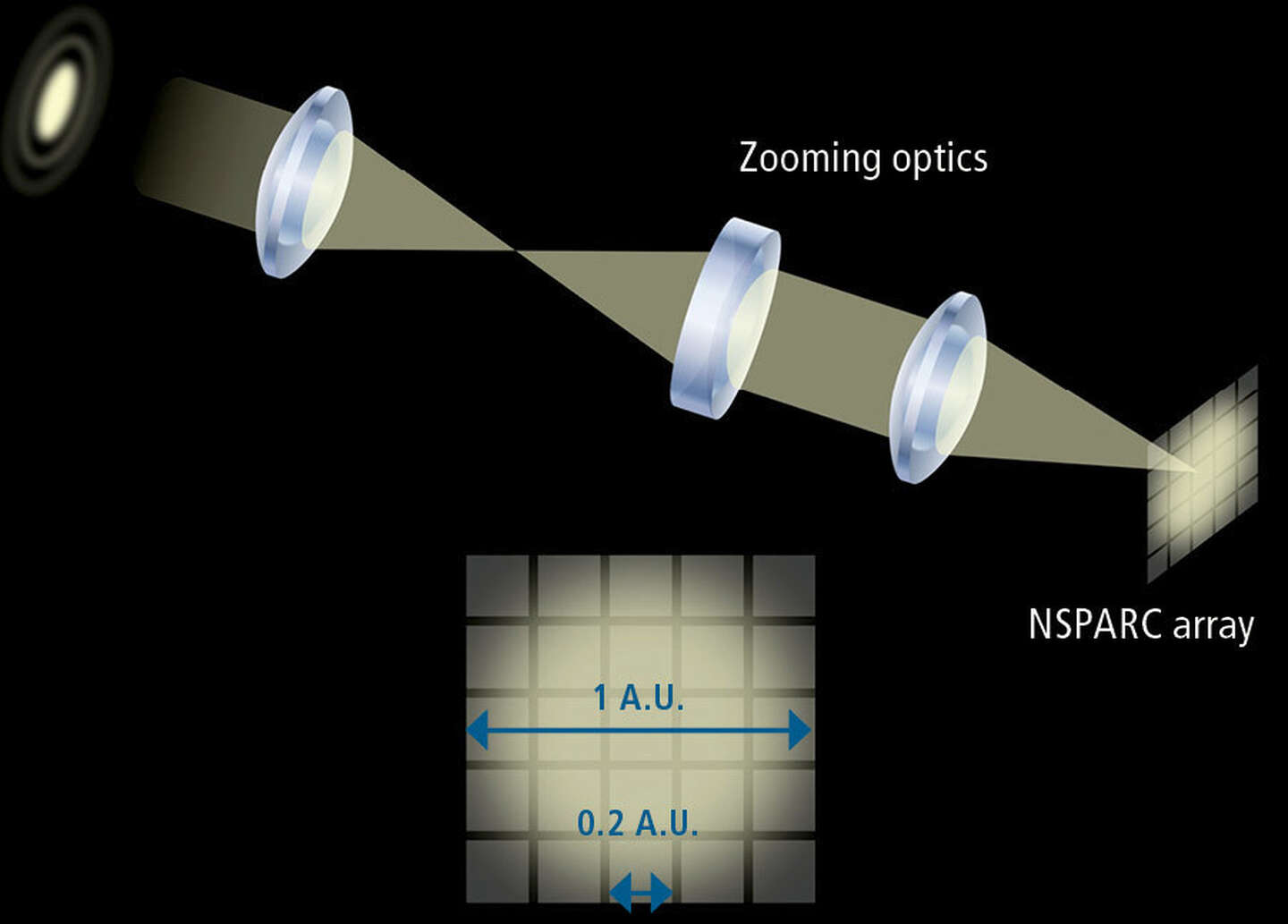 |
Each pixel from a traditional detector contains only one intensity value, and no spatial information.
|
| NSPARC
With NSPARC detection, the fluorescence emission light is directed through optics to the array detector, where the projected light fills the array.
|
AX/AX R
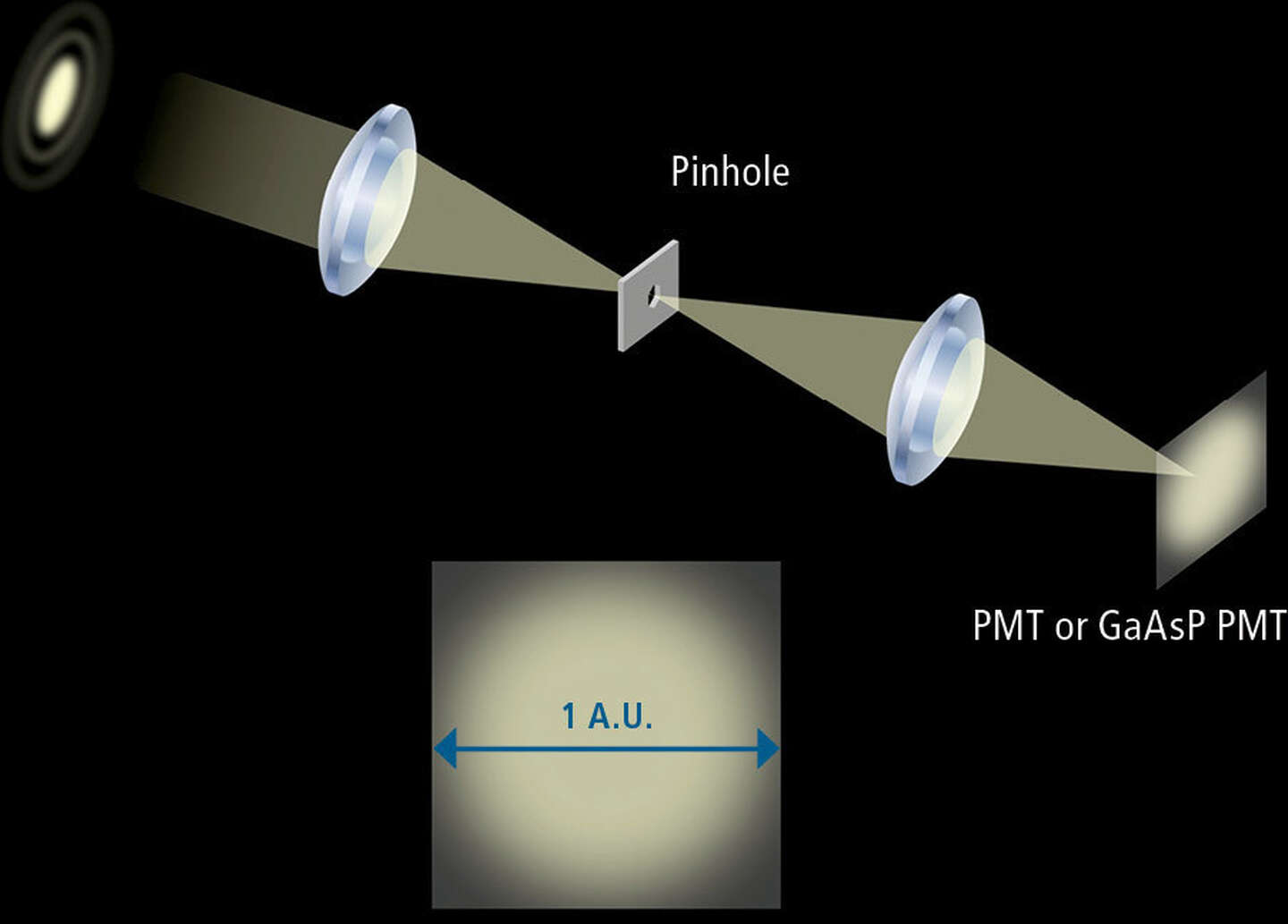
With traditional PMT or GaAsP detectors, the fluorescence emission light passes through a variable sized emission pinhole (typically set to 1 airy unit).
 NSPARC NSPARC |
 |
Traditional Detector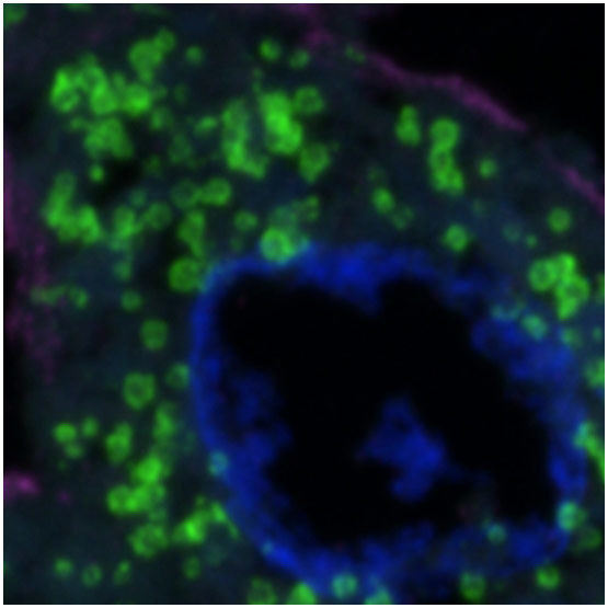
NSPARC
|
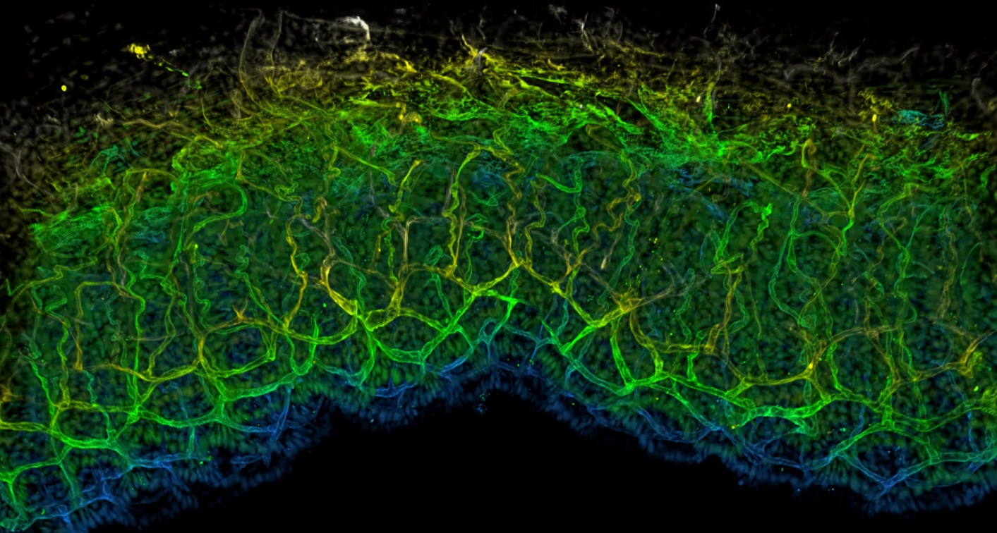
An Ideal Tool for Live Imaging Applications
The combination of ultra-short dwell times with the AX R resonant scanner and NSPARC’s sensitivity allows longer, less phototoxic imaging data to be collected in live-cell assays.
High Speed Imaging with Improved Sensitivity
With single photon detection sensitivity, the NSPARC detector’s extremely low noise profile and exceptional sensitivity make it a perfect match for AX R confocal resonant high-speed imaging, where the pixel dwell time is as short as 200 nanoseconds, enabling image acquisition at video frame rate. More details can be extracted from images for downstream analysis and computation.
Extremely Flexible Hardware Configurations
The NSPARC detector can be integrated on its own as the only detector for the AX/AX R, or as an additional detector for a multichannel AX/AX R system with either the DUX-VB or ST detector units and an optional transmitted light detector.
Current AX users can upgrade systems to add the NSPARC detector as well.
Superior Optics for Confocal
Controlling the manufacturing and implementation of optics from raw materials all the way to complete microscope systems brings unparalleled optical quality and performance.
Complementary optical design means the confocal system, microscope, and objectives all are optimized and matched for superior quality and resolution.
Nikon’s CFI60 and 75 infinity corrected optical system has numerous options for magnifications, working distances, and immersion mediums, paired for use with an extremely wide variety of samples and specimen preparations.
Nano Crystal Coat for superior transmission
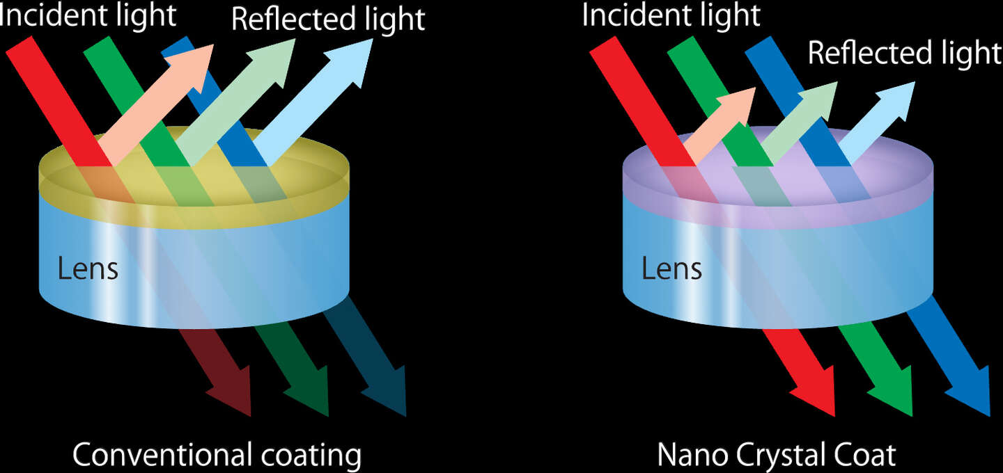
Nikon’s exclusive Nano Crystal Coat is an anti-reflective coating consisting of ultra-fine crystalline particles. This forms a coarse structure that lowers the reflectance index, facilitating the passage of light through the lens rather than reflecting it, and thus providing superior light transmission.
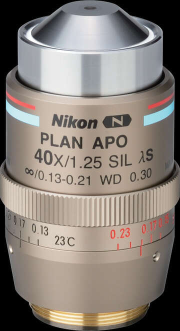
CFI Plan Apochromat Lambda S 40XC Sil
This objective provides bright, high-resolution images, and is suitable for imaging of thick samples and long-term time-lapse imaging.
| Working distance: | 0.30mm |
|---|---|
| Numerical aperture: | 1.25 |
| Chromatic aberration correction: | from visible to UV |
| Nano Crystal Coat applied | ✔︎ |
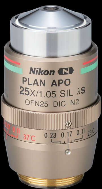
CFI Plan Apochromat Lambda S 25XC Sil
This objective provides a high NA, large FOV and long working distance, and uses silicone oil with a refractive index approximating that of live cells as the immersion liquid. It enables clear observation even in the deeper regions of thick samples.
| Working distance: | 0.55mm |
|---|---|
| Numerical aperture: | 1.05 |
| Chromatic aberration correction: | from visible to UV |
| Nano Crystal Coat applied | ✔︎ |
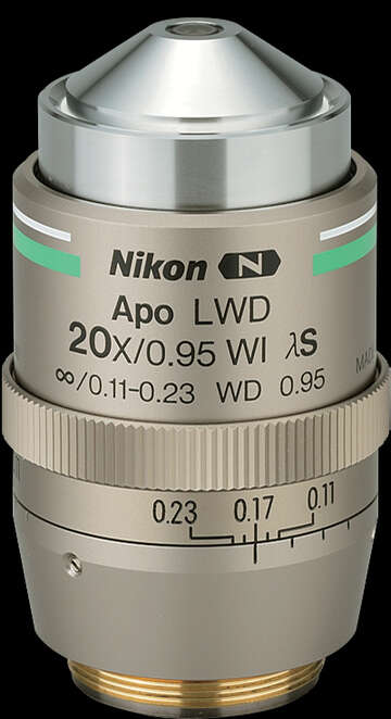
CFI Apochromat LWD Lambda S 20XC WI
This highly versatile objective provides superior performance thanks to its high numerical aperture, wide field of view and long working distance.
| Working distance: | 0.95mm |
|---|---|
| Numerical aperture: | 0.95 |
| Chromatic aberration correction: | from visible to infrared |
| Nano Crystal Coat applied | ✔︎ |
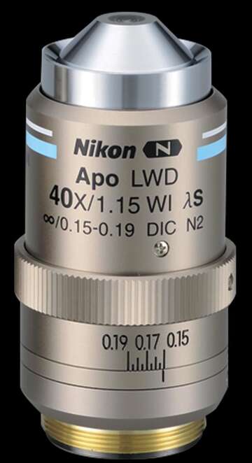
CFI Apochromat LWD Lambda S 40XC WI
Its high NA for 40X water immersion objectives provides brighter and higher-resolution images and makes this lens ideal for confocal live cell imaging.
| Working distance: | 0.6mm |
|---|---|
| Numerical aperture: | 1.15 |
| Chromatic aberration correction: | from UV through to near IR |
| Nano Crystal Coat applied | ✔︎ |
Objectives for confocal microscopy
| Model | NA | W.D (mm) | Nano Crystal Coat |
|---|---|---|---|
| CFI Plan Apochromat Lambda D 2X | 0.1 | 8.5 | ○ |
| CFI Plan Apochromat Lambda D 4X | 0.2 | 20.0 | ○ |
| CFI Plan Apochromat Lambda D 10X | 0.45 | 4.0 | ○ |
| CFI Plan Apochromat Lambda D 20X | 0.8 | 0.8 | ○ |
| CFI Plan Apochromat Lambda D 40X | 0.95 | 0.21 | ○ |
| CFI Plan Apochromat Lambda D 60X Oil | 1.42 | 0.15 | ○ |
| CFI Plan Apochromat Lambda D 100X Oil | 1.45 | 0.13 | ○ |
| CFI Plan Apochromat Lambda S 25XC Sil | 1.05 | 0.55 | ○ |
| CFI Plan Apochromat Lambda S 40XC Sil | 1.25 | 0.30 | ○ |
| CFI Plan Apochromat VC 60XC WI | 1.20 | 0.29 | – |
| CFI Plan Apochromat 10XC Glyc*1 | 0.50 | 5.50 | ○ |
| CFI Plan Apochromat Lambda 60XC | 0.95 | 0.15 | ○ |
| CFI Plan Apochromat IR 60XC WI | 1.27 | 0.17 | ○ |
| CFI SR HP Plan Apochromat Lambda S 100XC Sil | 1.35 | 0.30 (0.31-0.29): 23˚C, 0.29 (0.30-0.28): 37˚C |
○ |
| CFI Apochromat LWD Lambda S 20XC WI | 0.95 | 0.95 | ○ |
| CFI Apochromat LWD Lambda S 40XC WI | 1.15 | 0.60 | ○ |
*1: If you use a tissue clearing reagent, instead of glycerin, as an immersion liquid, some tissue clearing reagents may damage these products. Please confirm with a distributor before purchasing the product.