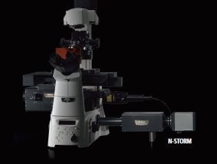


Super-resolution microscope system which exceeds traditional diffraction limits by an order of magnitude and is capable of 3D image acquisition.
Super-Resolution microscope system offering ten times the resolution of conventional optical microscopes.
N-STORM is a super-resolution microscope system that combines “STochastic Optical Reconstruction Microscopy” technology (licensed from Harvard University) and Nikon’s Eclipse Ti research inverted microscope. The N-STORM super-resolution microscope provides dramatically enhanced resolution that is 10 times that of conventional optical microscopes.
N-STORM utilizes a high accuracy localization information for thousands of individual fluorophores present in a field of view to create breathtaking super-resolution images exhibiting spatial resolution of 20nm; ten times greater than conventional optical microscopes.
In addition to lateral super-resolution, N-STORM utilizes proprietary methods to achieve a tenfold enhancement in axial resolution to 50nm, effectively providing 3D information at a nanoscopic scale

Single color 3D-STORM image of mitochondria in a BSC-1 cell labeled with Alexa405-Alexa647
Color encodes z-position information
Multi-color super-resolution imaging can be carried out using several dye strategies.
Unique tandem dye pairs (e.g. Alexa 405-Alexa 647, Cy2-Alexa 647, Cy3-Alexa 647) that combine “activator” and “reporter” probes allow highly accurate localization with no z shift.
An alternative dye strategy is to use standard commercially available secondary antibodies for continuous activation imaging to easily gain critical insights into the localization of multiple proteins at the molecular level.

The N-STORM system is based around the Nikon Ti-E inverted microscope. This flexible platform allows combination with confocals such as the A1R+ for live-cell imaging, low magnification observation, photo-stimulation and 20nm-resolution imaging on one integrated system, under the control of the universal NIS-Elements platform.


N-SIM and N-STORM can be combined on a single Nikon Ti-E inverted microscope to create the ultimate super-resolution imaging system. Using the N-SIM/N-STORM kit, switching between live cell super-resolution and 20nm-resolution imaging is possible without having to change the camera adapter.
|
Original Data |
Detected Molecules with |
Detected Molecules with |
| 351,560 mol.
Without FOP |
1,065,088 mol.
With FOP |
The SR Apochromat TIRF 100x 1.49 N.A. objective lens is designed to provide the highest quality point spread function for N-STORM and N-SIM super-resolution imaging

| Super-Resolution | Lateral (XY) ~20-30nm* Axial (Z) ~50nm* |
|---|---|
| 3D STORM Axial Range Per Single Data Set | ±500nm (up to 20 localized planes) |
| Microscope Platform | Ti-E TIRF with Perfect Focus System |
| Compatible Objectives | Plan Apo VC 100X 1.40 Oil Apo TIRF 100X 1.49 Oil |
| Available Lasers, AOTF Modulated | 405nm (20mW at the fiber tip) 457-514nm (70mW at the fiber tip) 561nm (70mW at the fiber tip) 647nm (125mW at the fiber tip) |
| Anti-Vibration Optical Table | Required |
| Camera | EM-CCD cameras iXon3 897, iXon Ultra 897 (Andor), and Evolve 512 Delta (Photometrics)**. Higher frame-rates can be achieved with the iXon Ultra and Evolve Delta. |
| Fluorescence Probe Combination (Photo-switchable STORM probes)*** | |
*Resolution depends on dye properties
**Camera compatibility depends on system configuration.
***Includes, but not limited to tandem-dye pairs
Super-resolution techniques have had a significant impact on our understanding of biological processes at the molecular level. However, one of the challenges to their broad utilization has been our limited ability to quantitatively analyze super-resolution images of complex biological tissues. In this Application Note, we highlight recent work by Dudok et al. utilizing Nikon’s N-STORM system to develop new correlative imaging methods and quantitative analysis tools to study the mechanism of cannabinoid signaling in the brain.
 Bioengineering
 Biomechanics
 Biophysics
 Cardiovascular Research
 Cell Biology
 Dermatology
 Developmental Biology/Embryology
 Hepatology
 Microbiology
 Molecular Biology
 Nephrology
 Neurobiology / Neuroscience
 Obstetrics & Gynaecology
 Oncology
 Ophthalmology
 Rheumatology
 Stem Cell & Regenerative Medicine
 Tropical Medicine
 Urology
 Vascular Research