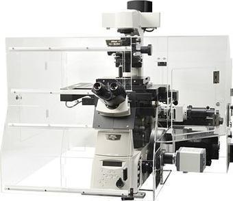
Scientists stand to find great value from the all-powerful 3D laser scanning confocal microscope which is breaking the limitations of imaging and is a must-have for a wide range of applications.
The ever-rising demand for advanced imaging solutions has resulted in the constant pursuit of a solution that will fill the gap in the scientific research field. Fortunately, the continually advancing technology which has been adopted in the industry has resulted in the ready availability of solutions which simplify day to day activities. A stiff neck competition by manufacturers and the high standards set by researchers have been the driving pillars which have redefined the solutions available in the market.
When it comes to imaging solutions, the 3D laser scanning confocal microscope is defying limits and bringing to hand endless possibilities. The super powerful microscope is everything that the research community desires and provides users with enhanced capabilities which include;
Improved confocal imaging resolution
A defining characteristic of the advanced microscopy solution is its high-resolution confocal imaging abilities. The microscope brings imaging to life thanks to its enhanced capabilities which are simply outstanding. By using a system of robust algorithms and GPU-based processing, there is no guesswork when it comes to de-convolution analysis.
Ultra-fast imaging
High-speed imaging abilities complement the top of the line confocal imaging resolutions of the microscope. Using either a resonant or Galvano scanner the microscope is able to achieve high frequencies which support the ultrafast image acquisition. Models which use the resonant scanner, in particular, are famous for producing the world’s fastest image acquisition as they are able to achieve a resonance frequency of 7.8 kHz. All this is complemented by a larger field of view which allows for the capturing of more details while maintaining top class image quality.
Advanced GaAsP Multi-Detector unit
Compared to a regular PMT, the GaAsP multidetector unit in confocal microscopes has far much better sensitivity which makes it possible for the acquisition of brighter signals even under weak fluorescence. An extra plus for the GaAsP PMT’s is they support all these functions with minimal background noise.
Real-time unmixing during image acquisition
The superior algorithms and superfast speeds of data processing by 3D laser scanning confocal microscopes enable for accurate spectral unmixing. This is made possible through the elimination of auto-fluorescence and separation of overlapping fluorescence spectra.
V-Filtering Function
There are different channels across different spectral ranges that can easily be selected to suit the spectrum of the fluorescence probe in use. Within each spectral range, filter-less intensity adjustment is supported which enhances the clarity of the super-resolution images.
Ease of use
The inclusion of user-centered software takes away a lot of guesswork when using the confocal microscope as it allows for integrated control through a more straightforward interface. All it takes to use the software is to have a control computer which meets the specifications which are clearly stated for all models. Since a confocal microscope has superior imaging capacities, they can be used for a whole line of applications including cell biology, biophysics, cardiovascular research, stem cell & regenerative medicine, neuroscience, and Ophthalmology among other research fields.