You might have heard the term 3D laser scanning confocal microscope. Also, when looking for microscopes you might have come across the words — high resolution and speed, 3D confocal images among others. But wait, what does the word “confocal” indicate and what is the microscopic system involved? And, how can it be applied whether you are into bioengineering, cell biology, or other related sectors? Well, read on to know more about the confocal microscope.
So What is Confocal?
Confocal” is described as “holding the identical focus.” What this implies in the microscope world is that the ultimate image has an identical focus or the focus resembles the point of focus in the target. The object and its final image are “confocal.” The modern-day confocal microscope can drain out the out-of-focus light from below and above the point-of-focus in the object. Usually, when an object or specimen is imaged in the conventional fluorescence or other forms of the microscope, the signal created is from the entire thickness of the object which does not let most of it be in focus to the beholder. On the contrary, the confocal microscope gets rid of this out-of-focus data through a confocal “pinhole” located in the foreground of the image plane which serves as a spatial filter and lets only the in-focus part of the light to be imaged. Hence, light from below and above the plane of focus of the specimen is excluded from the image.
Two Unique Elements that Come into Play
While the final image that is observed with a confocal microscope is all in-focus data, this generates another issue. In comparison to the conventional microscope, the quantity of light that is observed in the ultimate image is considerably decreased by the pinhole, generally up to 90%. To counterbalance for this lack of light, two unique elements have been included into modern day confocal microscopes. First of all, lasers are utilized as light sources in place of the traditional mercury arc lights since they offer notably bright light at very precise wavelengths for fluorochrome excitation. Secondly, extremely sensitive photomultiplier-detectors (PMTs) are engaged as imaging agents to pick up the diminished signal. The signal for detection in the modern-day confocal microscopes is generated by considering a concentrated laser beam over a square or rectangular range. A system of scanner mirrors sequentially examines a horizontal beam across the object.
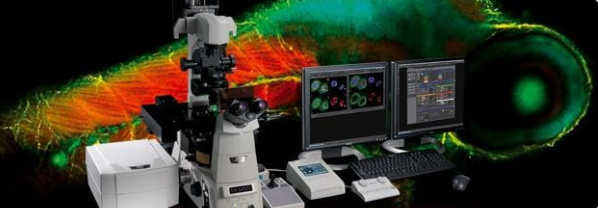
The Role of Computer Imaging
The third aspect that is fused into the confocal microscope is the latest microcomputer. It is accessed to command the scanner mirrors and the focusing system along with collect, save, and scrutinize the data. It can be saved in the form of digital pictures which may be seen on a computer monitor or it can be transferred to a hardcopy output device like a printer.
Computer imaging is an altogether different technology than direct photographic imaging. The computer allows the device to scan subsequent planes in the intended direction, save them, and build overlays of all the sections. This data can also be utilized to produce 3D images of the specimens. This gives you a complete view of the object on a microscopic scale in ways you have never imagined before. You can consider 3D laser scanning confocal microscope from a reputable supplier to achieve the best results.