New Supplier Announcement! – LaserAtWork GmbH

We are proud to announce that we have entered into a Distributor Agreement with LaserAtWork GmbH.

Founded in 2023, LaserAtWork GmbH mission is to provide customers with compact, cost-effective, and reliable ultrafast laser systems for medical applications, industry and science.

New Supplier Announcement! – Surface Optics Corporation

We are proud to announce that we have entered into a Distributor Agreement with Surface Optics Corporation.

Surface Optics Corporation (SOC) was founded in San Diego in 1976 by a group of skilled engineers who identified the need for an optical properties measurement lab to support Southern California’s burgeoning aerospace industry.

Drawing on their extensive experience from working on military aircraft, missiles, satellites, and Apollo mission projects at renowned companies such as Douglas Aircraft Company, Lockheed, North American Aviation, and Convair, SOC’s founders established a premier lab equipped with unique instrumentation.

New Supplier Announcement! – lightsource.tech GmbH

We are proud to announce that we have entered into a Distributor Agreement with lightsource.tech GmbH

lightsource.tech specializes in the development of professional, high-luminance light sources and other optical components and devices that meet the demanding requirements of industry and research and can be seamlessly integrated into existing systems and setups.

Andor CB2 sCMOS Series

Introducing the Andor CB2 sCMOS Series

Oxford Instruments is thrilled to announce the expansion of the Andor CB2 Series – a high speed, low noise sCMOS camera platform, designed to meet the needs of customers across the ultraviolet to the visible spectrum.

The CB2 Series now integrates five sensors, offering flexibility in speed, pixel size and resolution, while benefiting from Sony’s CMOS technology such as global shutter and low readout noise. One platform, three options, endless applications. Explore the CB2 Series from our website!


Tomocube Newsletter (Oct 2025)

Welcome to the latest edition of our newsletter, featuring a new Case Study conducted in collaboration with Okomera and the research team from Institut Gustave Roussy.

This study highlights how Tomocube’s holotomography technology, when integrated with Okomera’s droplet-based microfluidics, enables detailed, label-free analysis of chemotherapy response in patient-derived organoids (PDOs). Our approach provides quantitative insights into the complex architecture of heterotypic PDOs, distinguishing tumor–stroma organization and microenvironmental responses.


Figure 1 - Holotomography and Okomera microfluidics integrated workflow

Okomera’s droplet-based organoid culture combined with Tomocube holotomography and TomoAnalysis enables label-free, quantitative monitoring of drug responses in patient-derived organoid models.


Chemotherapy Response of Cancer Patient-Derived Tumoroids


🔬 Highlights from the study:

  • Drug sensitivity profiling: Okomera’s microfluidic platform enabled a reliable assessment of carboplatin sensitivity in Luminal B PDOs using only a minimal sample from a patient biopsy.

  • Heterotypic PDO characterization: Correlative imaging of HT and immunofluorescence reveals distinct cellular architectures, with both tumor cells and stromal-like compartments.

  • Tumor vs. stroma distinction: HT identified stromal compartments with higher refractive index (RI) granularity than tumor regions, suggesting a potential label-free marker of stromal presence.

  • Stromal modulation of drug response: Stroma-rich organoids displayed higher resistance against carboplatin treatment, consistent with the role of the tumor microenvironment in mediating therapy resistance.


These results highlight how combining microfluidics and HT provides a deeper insight into drug response mechanisms beyond conventional endpoint assays.

Figure 2 - Correlative imaging reveals tumor–stroma heterogeneity in biopsy-derived organoids.

Correlative imaging reveals tumor–stroma heterogeneity in biopsy-derived organoids. Immunostaining revealed heterogeneous tumor cell populations and stromal-like regions lacking epithelial markers (upper panel). HT further distinguished compartments, showing higher granularity in stromal-enriched regions (CK5/CK8-, pink box in lower panel) than in tumor-enriched regions (CK5/CK8+, blue box).


A Synergistic Workflow: Holotomography Meets Microfluidics

Tomocube holotomography: label-free 3D imaging and analysis

The HT-X1 series expands organoid research by providing universal, label-free imaging and quantitative analysis applicable to any PDO culture, revealing biophysical dynamics invisible to conventional assays.

By reconstructing 3D RI tomograms of live organoids without staining, it delivers:

  • Non-invasive, long-term imaging of optically thick organoids in flexible experimental design.

  • Quantitative biophysical readouts, such as dry mass, protein concentration, compactness, and granularity, for direct characterization of tumor-stroma heterogeneity and cell states.

  • AI-powered TomoAnalysis pipelines for reproducible segmentation and quantification—extensible to diverse organoid models and treatment studies.


Okomera microfluidics: high-throughput organoid culture

Okomera’s droplet-based microfluidic platform enables rapid, biopsy-ready PDO generation from as few as 30 cells. Within minutes, cells are encapsulated into droplets that form uniform organoids, enabling:

  • High-throughput multiplexed screening with barcoded drug libraries (O-Plex).

  • Fast turnaround, with compact 3D organoids formed within 48 hours.

  • Automated AI-driven analysis of growth, viability, and dose–response curves.

This streamlined approach allows researchers to maximize limited patient material while testing a wide range of drug conditions in parallel.

By integrating Tomocube’s HT and Okomera’s microfluidic technology, researchers can combine high-throughput screening with deep, label-free imaging—uncovering drug resistance and tumor heterogeneity that conventional methods often miss.


Download the full case study

Tomocube Launches the HT-X1™ mini – Holotomography: Now for Every Lab


Daejeon, South Korea — [September 10, 2025] — Tomocube Inc., a leading innovator in holotomography (HT) technology, has announced its new HT imaging system, HT-X1™ mini. This product has been designed to make advanced HT technology more accessible to researchers, aiming to expand the adoption.

The HT-X1 mini delivers high-resolution 3D HT imaging comparable to Tomocube’s HT-X1 and HT-X1 Plus models. The system can be upgraded by integrating additional modules such as 3D fluorescence, stage-top incubator, multi-wavelength holotomography optics, etc. Researchers tailor the system to their specific needs, enhancing its accessibility and strengthening the company’s competitive edge in diverse applications.

The data analysis is fully compatible with TomoAnalysis™, the image analysis software supporting seamless AI-powered analysis, marking a transformative step in delivering advanced scientific tools that are both powerful and accessible.

This strategic development will allow researchers to leverage advanced HT technology with greater accessibility. The HT-X1™ mini, with its compact design, empowers researchers with enhanced usability in everyday experiments. By combining affordability with performance, it enables the seamless integration of advanced 3D live-cell imaging into routine laboratory workflows.

With this launch, Tomocube has established a complete product lineup from entry-level to flagship systems. This comprehensive range aims to broaden researcher accessibility across diverse fields including organoids, cell therapy, drug development, and in vitro fertilization (IVF) embryo assessment, while accelerating the adoption of HT technology in global research environments.

“The HT-X1 mini lowers barriers to entry in research settings, allowing more researchers to experience our 2nd-generation holotomography technology,” said Prof. YongKeun Park, CEO of Tomocube. “With this launch, we aim to enable more scientists to experience its benefits, while continuing to provide the highest standards of product quality and customer support.”

The launch of the HT-X1™ mini represents a significant milestone in Tomocube’s mission to deliver advanced, non-invasive imaging tools that empower researchers and accelerate scientific discovery across diverse fields of life science.  Moving forward, Tomocube plans to actively pursue global expansion beyond North America and Europe into new markets including Asia and South America, while strengthening its comprehensive one-stop support system covering installation, training, and software analysis to accelerate worldwide adoption.

Tomocube Newsletter (Oct 2024)

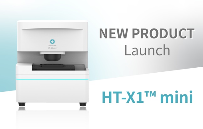
Transforming 3D Biology Using AI: Tomocube’s HT-X1™ Plus Accelerates Cellular and Organoids Label-Free Analysis

Tomocube, a global leader in 3D label-free imaging and analysis, proudly introduces the HT-X1 Plus, a state-of-the-art bioimaging platform designed to meet the evolving needs of biomedical researchers. This new system raises the bar in high-resolution, high-throughput 3D imaging for cells and organoids, providing researchers with faster, more detailed, and more accurate insights into biological processes.

With the HT-X1 Plus, Tomocube has integrated cutting-edge technologies that enhance precision, imaging speed, and data richness. The platform offers a 4x larger field of view and advanced illumination optics, making it a game-changer for phenotypic screening, tissue section imaging, and the study of fast-moving microorganisms. These capabilities unlock new research possibilities in cell biology, regenerative medicine, and organoids research.


Setting New Standards in 3D Imaging

Building upon the proven success of the HT-X1, the HT-X1 Plus sets new standards in 3D imaging performance. Equipped with a high-performance CXP camera and AI-powered image reconstruction algorithms, the platform drastically reduces scan times, enabling comprehensive 3D imaging of a full 96-well plate in under 30 minutes—without sacrificing detail or quality. “With the HT-X1 Plus, researchers can now explore complex biological samples like organoids with unparalleled clarity and speed,” said YongKeun (Paul) Park, Chief Executive Officer in Tomocube. “This platform is designed for high-content, image-based drug screening assays and holds tremendous potential to accelerate discoveries in biomedical research.”


Key Features of the HT-X1 Plus Include:

* Larger Field-of-View: Capture expansive areas without stitching, ideal for large-scale, high-content experiments.
* Faster Image Acquisition: Optimized for high-throughput screening, capable of scanning 96-well plates in just 30 minutes.
* Flexible Light Source Options: Configure imaging with three wavelengths (R/G/B) for optimal contrast and penetration.
* Advanced Correlative Fluorescence Imaging: Integrated sCMOS-based fluorescence module (FLX) enhances 3D imaging with high sensitivity and precision.
* Color Brightfield Imaging: A new modality with wide preview scan mode improves histological studies, providing comprehensive tissue morphology analysis.


Solving Major Research Challenges:

The HT-X1 Plus directly addresses key challenges in bioimaging, including slow scan speeds, limited fluorescence sensitivity, and challenges related to light absorption in thick samples. Its advanced imaging capabilities—such as reduced phototoxicity and improved 3D optical sectioning—make it the ideal tool for investigating dynamic, sensitive samples without compromising image quality.


Pushing the Boundaries of Biomedical Discovery:

Designed to integrate effortlessly with molecular studies and fluorescence-tagged sensors, the HT-X1 Plus enables researchers to visualize and quantify intricate biological processes with unprecedented detail. The platform’s AI-driven TomoAnalysis software facilitates in-depth analysis, further pushing the boundaries of cellular dynamics research.


Applications of HT-X1 Plus Include:

* Cell Biology & Regenerative Medicine: Perform high-content screening of live cells and organoids for drug discovery and therapeutic studies.
* 3D Biology & Organoids: Obtain detailed imaging of dense organoids, tissue sections, and immune cell interactions.


Tomocube Newsletter (Sept 2024)

Holotomography Featured on the Cover of Nature Reviews Methods Primers

In the rapid evolving world of Holotomography (HT), its potential applications are nothing short of breathtaking (and yes, many Tomocube users have, quite literally, found themselves breathless at times.) From biological research to cutting-edge medical diagnostics, HT is transforming the way we see the world at the microscopic level.

While the future is thrilling, sometimes it’s essential to step back and revisit the fundamentals that make the technology so revolutionary.

To truly grasp the depth and impact of HT, we encourage you to dive into a comprehensive review, “Holotomography”, recently published in the prestigious Nature Reviews Methods Primers. Being selected for the cover of July’s Primer is a testament to the groundbreaking impact of Holotomography.

This edition will provide an overview, but the article offers detailed insights that are essential for anyone looking to stay at the forefront of advancements in biomedical imaging.


Holotomography – the Technological Innovations it Brings to the Field

Key highlights of the article:

  • *Revolutionary Technology: Discover the inner workings of HT! Gain insights into how its precision, adaptability, and label-free imaging capabilities can enhance your research, offering a powerful alternative to traditional microscopy with superior clarity and quantitative analysis.


  • *Wide Applications: An exploration of HT wide-ranging applications, from basic research to advanced medical diagnostics. It’s not just about the technology itself, it’s about how HT can be integrated into your work to drive new understanding and breakthrough.


  • *Future Trajectories: A forward-looking perspective on the bright future ahead. This includes potential innovations and the integration of HT with emerging technologies such as regenerative medicine, 3D biology, and organoid-based drug discovery.


  • *Challenges and Optimization: A balanced discussion of HT’s strengths and current limitations, offering insights into potential optimizations and future improvements to equip you with the knowledge to navigate these challenges.

This article on HT is more than just a read—it’s a gateway to a transformative technology that could redefine your approach to cellular imaging and analysis. Dive in now and discover the potential that HT holds for your research!

New Supplier Announcement! – Opto

We are proud to announce that we have entered into a Distributor Agreement with Opto.

Opto GmbH has been developing and manufacturing compact imaging modules for industry and medical technology for 40 years.

Opto supports device developers and OEM’s in the development of their optical requirements – from prototype to series production.

New Supplier Announcement! – Telops Inc

We are proud to announce that we have entered into a Distributor Agreement with Telops Inc.

Telops designs and manufactures high-performance hyperspectral imaging systems and infrared cameras for defence, industrial, and academic research applications.

Telops also offers R&D services for optical systems technology development in order to respond to the specific needs of its customers.

Since its beginnings in 2000, Telops has distinguished itself with the quality of its personnel and its innovative approach to the technological challenges of the optics and photonics field. Today, the expertise of its scientists, engineers and technicians and the performance of its infrared cameras and hyperspectral imagers are internationally recognized.

While being headquartered in Canada, Telops caters to an international market using an efficient network of distribution and representation.