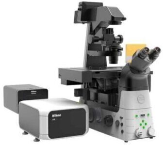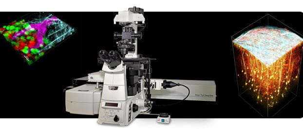
Streamlined multiphoton confocal for deeper, faster multiphoton imaging.
Nikon’s A1 MP+ is a streamlined version of the unique A1R MP+ multiphoton imaging system developed for simplified, cost-effective multiphoton imaging.
Nikon’s A1 MP+ multiphoton confocal microscope is a unique multiphoton imaging system featuring a high resolution galvanometer scanner. New 4 channel non-descanned detectors with higher sensitivity, reduced dark current and broad spectral range allow for real time unmixing of closely spaced probes for accurate and high-contrast spectral imaging. This is especially important in multiphoton microscopy because of the overlap of emission spectra of probes and autofluorescence, which is often unavoidable when using a single laser line.
The fluorescence emissions from deep within a specimen are highly scattered in multiphoton excitation, and therefore the conventional detector using a pinhole cannot provide bright fluorescent images. The episcopic NDD in the A1R MP+ is located close to the back aperture of the objective to detect the maximum amount of scattered emission signals from deep within living specimens.
The cerebral cortex of an H-line 5-week-old mouse was studied with the open skull method. The entire shape of dendrites of pyramidal cells in layer V expressing EYFP were visualized from the bottom layer into a superficial layer. In addition, the fluorescence signal of white matter in deeper areas was also studied.

| Left | 3D reconstruction image | Photographed with the cooperation of:Dr. Tomomi Nemoto, Research Institute for Electronic Science, Hokkaido UniversityDr. Shigenori Nonaka, National Institute for Basic BiologyDr. Takeshi Imamura, Graduate School of Medicine, Ehime University |
|
| Right | Z-stack images | ||
| Top: dendrites located in superficial layers in the layer V pyramidal cells 25 µm from the surface |
|||
| Middle: basal dendrites in the layer V pyramidal cells 625 µm from the surface |
|||
| Bottom: fluorescence from white matter Excitation wavelength: 950 nm Objective: CFI75 Apo 25xW MP (NA 1.10 WD 2.0) |
Mouse cerebral cortex multi-color imaging
 |
 |
 |
Simultaneous acquisition of three channels in anesthetized YFP-H mouse using IR excitation of 950 nm and imaging Second Harmonic Generation (SHG) and two fluorescence emissions.
Cyan: SHG signal of dura mater
Yellow: EYFP pyramidal neurons in layer V of the cortex
Red: SRB-labeled blood vessels
Photographed with the cooperation of:
Drs. Ryosuke Kawakami, Terumasa Hibi and Tomomi Nemoto, Research Institute for Electronic Science, Hokkaido University
3D volume rendering images
Three-dimensional volume renderings of a kidney labeled with Hoxb7/myrVenus marker (Chi et al, 2009 Genesis), using depth-code pseudocolor volume rendering to reference Z depths (pseudocolored by depth – 1 μm step for 550 μm).

Objective: CFI Apochromat 25xW MP, Scan zoom: 1x, Z step size: 1 μm, IR excitation wavelength: 930 nm
Image resolution: 1024×1024 pixels, Image volume: 460 μm (length) x 460 μm (width) x 600 μm (height)
Photographed with the cooperation of Dr. Frank Costantini and Dr. Liza Pon, Columbia University Medical Center, New York
In addition to the GaAsP NDD compatible with a wavelength of 1080nm, there is a new model for upright microscopes that is compatible with a wavelength of 1300 nm. This new NDD enables deep imaging up to 1.4 mm in combination with a newly developed scanning head A1R MP+ that is compatible with a wavelength of 1300 nm.
Deep brain imaging in in vivo mouse with the GaAsP NDD compatible with the 1300 nm wavelength

In vivo imaging of an anesthetized YFP-H mouse (4-week-old) via open skull method. Visualization of the entire layer V pyramidal neurons and the deeper hippocampal neurons. Deep imaging achieved for 3-dimensional imaging of hippocampal dendrites up to 1.4 mm into the brain.
Captured with episcopic GaAsP NDD for 1300 nm and CFI75 Apochromat 25xW MP1300 objective lens (NA 1.10, WD 2.0 mm)
Excitation wavelength: 1040 nm
Mouse brain in vivo dual color imaging with the GaAsP NDD compatible with the 1300 nm wavelength

The cerebral cortex of an anesthetized YFP-H mouse (4-week-old) was studied with the open skull method.
Alexa594 was injected into the tail vein to visualize the blood vessel.
High-NA objectives have been developed that highly correct chromatic aberrations over a wide wavelength range, from ultraviolet to infrared. Transmission is increased through the use of Nikon’s exclusive Nano Crystal Coat technology.
In particular, the CFI75 Apochromat 25xW MP/MP1300 objective lenses provide an industry leading highest numerical aperture of 1.10 while still maintaining a 2.0 mm working distance. They also have a collar that corrects spherical aberrations depending on the depth of the specimen and a 33° manipulator pipette access angle, making it ideal for deep multiphoton imaging and physiology research applications.
Nano Crystal Coat is a Nikon exclusive lens coating technology using an ultralow refractive index nanoparticle thin film originally developed for the semiconductor fabrications industry. The Nano Crystal Coat particle structure dramatically reduces stray reflections and boosts transmission over a wide wavelength range, producing images with higher signal-to-noise (S/N) ratios.

CFI75 Apochromat 25xW MP CFI Apochromat LWD 40xWI λS

CFI Apochromat 40xWI λS CFI Plan Apochromat IR 60xWI

CFI Apochromat LWD 20xWI λS
| CFI75 Apochromat 25xW MP1300 |
NA 1.10 WD 2.0 Nano Crystal Coat |
|---|---|
| CFI75 Apochromat 25xW MP |
NA 1.10 WD 2.0 Nano Crystal Coat |
| CFI Apochromat LWD 20xWI λS |
NA 0.95 WD 0.95 Nano Crystal Coat |
| CFI Apochromat LWD 40xWI λS |
NA 1.15 WD 0.6 Nano Crystal Coat |
| CFI Apochromat 40xWI λS |
NA 1.25 WD 0.18 Nano Crystal Coat |
| CFI Plan Apochromat IR 60xWI |
NA 1.27 WD 0.17 Nano Crystal Coat |
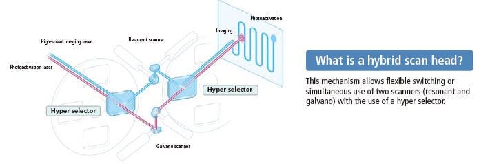
When the multiphoton laser wavelength or group velocity dispersion pre-compensation is changed, the multiphoton laser beam positional pointing at the objective back aperture may also change, resulting in uneven intensity across the image, or a slight misalignment between the IR and visible laser light paths.
Verifying the IR laser beam pointing and setting the alignment has traditionally been difficult. The A1R MP+ auto laser alignment function, housed in the Incident Optical Unit for the multiphoton excitation light path, automatically maximizes IR laser alignments with a single click in NIS-Elements C.
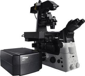
Streamlined multiphoton confocal for deeper, faster multiphoton imaging.
Nikon’s A1 MP+ is a streamlined version of the unique A1R MP+ multiphoton imaging system developed for simplified, cost-effective multiphoton imaging.
Nikon’s A1 MP+ multiphoton confocal microscope is a unique multiphoton imaging system featuring a high resolution galvanometer scanner. New 4 channel non-descanned detectors with higher sensitivity, reduced dark current and broad spectral range allow for real time unmixing of closely spaced probes for accurate and high-contrast spectral imaging. This is especially important in multiphoton microscopy because of the overlap of emission spectra of probes and autofluorescence, which is often unavoidable when using a single laser line.

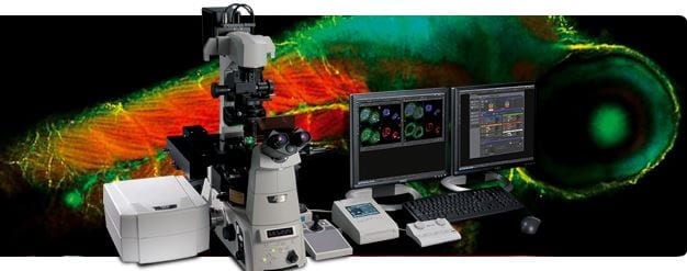
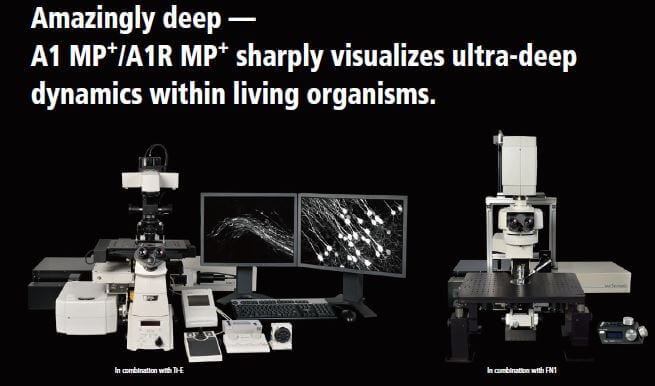
Ultrafast resonant scanners; high-sensitivity, low-noise GaAsP detectors; and industry-leading optics have made Nikon’s A1R MP multiphoton confocal microscope a system of choice for deep tissue imaging. Recent collaborative efforts between Nikon and the Allen Institute for Brain Science resulted in the design of an open-architecture multiphoton system able to image large specimens. Stimulated by this collaboration, Nikon has developed a commercially available large-format multiphoton system that can be used for the most demanding intravital imaging applications.
 Cardiovascular Research
 Cell Biology
 Dermatology
 Hepatology
 Molecular Biology
 Nephrology
 Neurobiology / Neuroscience
 Obstetrics & Gynaecology
 Odontology (dentistry)
 Oncology
 Orthopaedics
 Stem Cell & Regenerative Medicine
 Tropical Medicine
 Urology
 Vascular Research