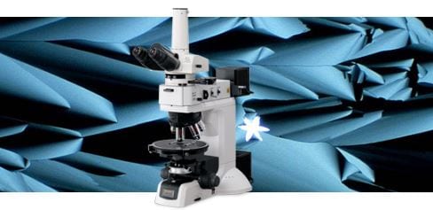
Y-CAM high resolution wireless camera

Y-CAM is an easy to operate compact size camera with built in battery and Wi-Fi generator which allow multiple users to view the same live image simultaneously.

Y-CAM high resolution wireless camera

Y-CAM can be easily installed into most of the microscope and immediately upgrade into WIFI electronic eyepiece. It allow multiple viewers to view it at the same time through IOS or Android devices with the aid of Ucam plus application.

Features

– Built in 5 megapixel color sensor
– Built in wireless network
– IOS and Android compatibility
– Up to 10 viewers at the same time

Bioptechs – FCS2 System

The FCS2® System is a closed system, parallel plate flow cell environmental chamber used on a microscope to enable the acquisition of high resolution, time-lapse images of cells undergoing either natural growth or fluidic exposure to variant factors. The FCS2® System is designed to be used on any inverted microscope as the FCS3® System is specifically for upright microscopes.

Bioptechs – FCS2 System

The Focht Chamber System 2 (FCS2®) is a closed system, live-cell micro-observation chamber that utilized several patented technologies that offer several exclusive advantages over other chambers. In addition to its uniform temperature control and user definable perfusion capability, it is fully compatible with all modes of microscopy. It is also the only chamber to combine high-volume laminar flow perfusion rates with Koehler illumination and precise temperature control without the need for an air curtain. This is accomplished by the development of micro aqueduct perfusion. A method of introducing media into a fluid optical cavity where the separation of optical surfaces and flow geometry is definable by the user. Therefore, the chamber is adaptable to the protocol instead of having to adapt the protocol to the chamber.
 
The FCS2 is a flow cell or flow chamber that is designed specifically for the demands of today’s live-cell imaging requirements. It has limitless flow characteristics because its flow geometry can easily be customized by the user. It provides the ability to configure the optical cavity to produce control shear forces, flow characteristics, high or low volume exchange rates, near laminar flow, and anything in between. The most important feature of the FCS2, other than unequalled temperature uniformity, is that the is flow of media is constrained to a precise location over the cells. Open dish type chambers do not provide the flow capabilities of the FCS2. The FCS2 provides unmatched, uniform temperature control, in a user configurable, perfusable, optical imaging cavity.

FCS2 users enjoy the following characteristics:

  • Easy no tools needed assembly
  • Compatibility with all modes of microscopy
  • Complete control of the volume within the optical cavity
  • Complete control of the separation between optical surfaces
  • Complete control of the shape or profile of the flow channel
  • Complete control of the temperature uniformly across the entire field
  • Compatible with inverted microscopes

How does it work?

Cells are grown on a 40mm glass coverslip. This coverslip is then incorporated into a perfusable fluid optical cavity that is compatible with all modes of microscopy, and its geometry can be easily defined by the user. This optical cavity is secured into a fixture on the stage of the microscope where it can be perfused with media or remain static. Media that comes into one of the ports on the side of the chamber, emerges in a fluid optical path where the media is precisely directed over the cells. The media is collected within the optical cavity and directed out of the chamber on the other side. The flow characteristics of the media while in the optical cavity are easily modified by selecting gaskets of varying geometry that direct the flow. Temperature control is maintained by both peripheral thermal support from the chamber’s metal frame and heat that is emitted from an electrically conductive optically transparent coating on the “window” of the optical cavity. This technique provides uniform temperature control across the entire field, thus eliminating the temperature gradient that occurs with other designs. This technique also enables the FCS2 to recover from perfusion induced temperature fluctuations in just seconds.

Temperature Control:

The FCS2 was designed to maintain accurate thermal control and allow high-volume laminar flow perfusion. Both of these functions are incorporated into our patented Microaqueduct Slide (see drawing below). The surface of the slide, opposite the specimen side, is coated with an electrically conductive transparent thin film of Indium-Tin Oxide (ITO) and two electrical contacts (busbars). When the FCS2 is completely assembled two electrical contacts, (not shown in drawing), which are contained in the electrical enclosure rest on the busbars. A temperature controller is used to pass a regulated current flow through the ITO Coating. This causes the surface of the slide to heat. The heat is transferred through the perfusable media to the cell surface on the coverslip thereby providing first surface thermal control. The self-locking base of the chamber is also temperature regulated to provide peripheral heat as well.

Thermographic Images:

vislightfcs2
fcs237deg

The thermograph above demonstrates the uniform temperature distribution of an FCS3. Notice that the coverslip temperature is so uniform that its location, in infrared, is indistinguishable from the base of the chamber. This demonstrates the effectiveness of the ITO heated Microaqueduct Slide. It is capable of re-equilibrating cell temperature within seconds of perfusion and eliminates the typical thermal gradient that occurs with peripheral heating.

Microaqueduct Perfusion:

A fluid pathway is formed by separating the Microaqueduct Slide from the coverslip containing cells with a single silicone gasket. This gasket can be any thickness from 50 micron to 1mm and any lateral geometry you choose or create. This arrangement allows the user to define the flow characteristics. Therefore, you are not limited by the geometry of the optical cavity. Instead you select or create it! Fluid access to this flow channel is made through two 14-gauge needle stock tubes protruding from the sides of the chamber top. These tubes provide fluid connection to two perfusion holes in the Microaqueduct Slide that interface two “T” shaped grooves cut into the inner surface of the Microaqueduct Slide. The “T” groove allows the media to seek the path of least resistance and become nearly laminar before flowing across the cells. This technique eliminates the need for the metal perfusion ring and additional gaskets, which are the limiting factors, required by most conventional chambers. (see drawing below)

microaquflo microaqfcs2

Isometric View of Optical Cavity

Microaqueduct Slide is transparent glass but rendered as a solid to improve contrast to illustrate function. Microaqueduct design enables proper Koehler illumination with high-numeric aperture optics for both transmitted and reflected modes of microscopy. Suitable for static or high rate of flow procedures where a rapid exchange of media is required with low cell surface shear Cell temperature can be controlled from ambient to 50 degrees C +/- 0.2 degrees C without the need of an air curtain. Temperature is controlled uniformly across entire field with media equilibrating as it enters the chamber. (see drawing above)

XY Linear Motor Stages for Inverted Microscopes – HLD117

Prior’s newest stage technology, the HLD117 Stages with Linear Motor Technology brings a new level to precision in microscope automation.

XY Linear Motor Stages for Inverted Microscopes

The HLD117 series of flat top stages with linear motor technology for inverted research microscopes set new standards for convenience and performance. Designed to seamlessly integrate with other items such as micromanipulators and incubation chambers, the top surface of the stage is completely free of obstruction. This provides a highly ergonomic platform with exceptionally easy access to the sample area. The HLD117 incorporates constant contact bearings giving the lowest friction and smoothest drive available. 50 nm encoders are integrated into the stage as standard.

The HLD117 is:

Ultra Quiet

Even at high speeds (up to 300 mm/sec) the linear DC servo motors are almost silent.

Fast and Precise

With a maximum speed of 300 mm/sec and a minimum speed of 1 µm/ sec the HLD117 is suitable for a wide array of applications, and accerleration is so smooth imaging can be performed as the stage moves. The stage is highly precise, with 0.15 µm repeatability.

Low Profile

The low profile flat top design allows ample room to access microscope adjustments.

Easy to integrate

The slide is controlled via the ProScan III system, offering superlative control and the potential to automate much of the imaging process. With the addition of illumination devices, the Nanopositioning Piezo Z stage, and a robotic loader, one gains an extremely powerful system, with tight control over illumination, slide loading, and all three axes – ideal for high throughput applications where both speed and precision are vital. The stage is also compatible with third party software, and a software development kit is supplied with the stage to allow easy integration.

The HLD117’s offer superior versaltility and are an ideal solution for:

Slide Scanning

Built for performance with unmatched acceleration rates and speeds, the HLD117 is the best choice for either start/stop point to point imaging or for continuous motion and velocity controlled scanning applications. The ProScan controller with TTL I/O and triggering functions provides constant speed movements with extremely low velocity ripple for picture perfect synchronization between automation and imaging software.

Multipoint Imaging

Whether your cells are fixed or in solution Prior linear drive stages make multiple position imaging both rapid and precise. Image acquisition from multiple points scattered across a wide area is faster than previously thought possible – a 96 well plate can be scanned in approximately 15 seconds. The addition of Prior’s Nanopositioning Piezo Z stage, offering precise focussing for high end applications, allows three dimensional data collection in miliseconds.

XY Microscope Stages for Upright Microscopes – H101A

An extensive range of motorized XY stages to fit the majority of upright microscopes available.

XY Stages for Upright Microscopes – H101A

The H101A ProScan™ stage is adaptable to virtually any microscope or optical system. The H101A stage enables the user to increase labor efficiency by automatically scanning your specimens and storing points for later recollection and inspection with unmatched repeatability. The H101A is ideal for scanning or inspection of slides, microtitre trays, semiconductor wafers, or metallurgical specimens in any sort of pattern, including raster, snake and random patterns. The H101A ProScan™ stage also incorporates the patented – Intelligent Scanning Technology (IST). In conjunction with extensive testing, Intelligent Scanning Technology (IST) allows each stage to be pre-programmed with a unique set of operating characteristics particular to that stage to ensure optimum performance. IST (also available on H107 and H117 models) allows the ProScan™III controller to make any required adjustments to maintain superior orthogonally and metric accuracy.

XY Microscope Stages for Inverted Microscopes – H117

An extensive range of motorized XY stages to fit the majority of inverted microscopes available.

XY Microscope Stages for Inverted Microscopes – H117 for current inverted microscopes (114mm x 75mm)

The H117 stage series has a completely flat top for easy access, and includes Intelligent Scanning Technology (IST). In conjunction with extensive testing, IST allows each stage to be pre-programmed with a unique set of operating characteristics particular to that stage to ensure optimum performance. IST allows the ProScan™III controller to make any required adjustments to maintain superior orthogonality and metric accuracy.

Click here for datasheets about  stages for Nikon TE2000 microscopes, here for Nikon Ti microscopes,here for Olympus microscopes, here for Zeiss microscopes and here for Lecia microscopes. They have been used for cancer research in a system for obtaining extremely high quality images.

XY Microscope Stages for Inverted Microscopes – OptiScan ES107

An extensive range of motorized XY stages to fit the majority of inverted microscopes available.

XY Microscope Stages for Inverted Microscopes – OptiScan ES107 for inverted microscopes (114mm x 75mm)

The ES107 OptiScan stage for inverted microscopes offers the optimum compromise between price and performance. The ES107 has been designed to utilise the range of specimen holders from Prior’s ProScan stages. This enables examination of the widest range of specimen types including glass slides, multi-well plates, Petri-dishes and polished metallurgical samples.

XY Microscope Stages for Inverted Microscopes – H139 Multi-Well Plate Motorised Stage

An extensive range of motorized XY stages to fit the majority of inverted microscopes available.

XY Microscope Stages for Inverted Microscopes – H139 Multi-Well Plate Motorised Stage

Offering precise movement (repeatability 0.7 µm) in the X and Y axis (280 x 80 mm), the H139 is the perfect tool for labs tasked with precise microscopic examination of larger numbers of microplates or microscope slides, and gives users a uniquely powerful combination of precision, speed and convenience.

Designed for use with leading models of inverted microscopes, the H139 allows users to conduct two live cell imaging experiments in mircoplates at the same time, allowing more efficient use of imaging centre resources. Incorporating Prior’s patented Intelligent Scanning Technology (IST) as standard, the H139 can be optionally equipped with 50 nm encoders for demanding applications requiring the finest precision and accuracy.

XY Microscope Stages for Upright Microscopes – H101F Flat Top Stage

An extensive range of motorized XY stages to fit the majority of upright microscopes available.

XY Stages for Upright Microscopes – H101F Flat Top Stage

Prior Scientific proudly introduces their latest and most innovative motorized stage for upright microscopes, the H101F flat top stage. Significant benefits of the new ProScan H101F include a completely flat top plate design that provides unlimited obstruction free space for objective and nose piece rotation. Ultra lowprofile sample holders facilitate the use of high NA objectives/condensers while the latest in miniature high torque motors facilitate access to the condenser and other microscope adjustments. The larger sample holder design area accommodates a variety of applications such as large tissue sections and other custom sample holders. Optional embedded high resolution XY encoders provide the highest precision available. The H101F features smaller gauge stage cables which simplify cable routing. Older sample holders are also compatible with the new H101F by adding a simple adapter plate. The H101F ProScan™ stage incorporates the patented – Intelligent Scanning Technology (IST). In conjunction with extensive testing, Intelligent Scanning Technology (IST) allows each stage to be pre-programmed with a unique set of operating characteristics particular to that stage to ensure optimum performance. IST (also available on H101A, H107 and H117 models) allows the ProScan™III controller to make any required adjustments to maintain superior orthogonally and metric accuracy.

XY Microscope Stages for Upright Microscopes – H105 for Semiconductor Wafer Inspection

Product Details

An extensive range of motorized XY stages to fit the majority of upright microscopes available.

XY Stages for Upright Microscopes – H105 for Semiconductor Wafer Inspection (154mm x 154mm)

The H105 is especially well-suited for applications that typically involve large specimens. For example, for performing scanning of a wide range of semiconductor wafers, photo masks, and printed circuit boards. The H105 can easily accommodate 6″ wafers. H105 stage are fully mapped for improved metric accuracy