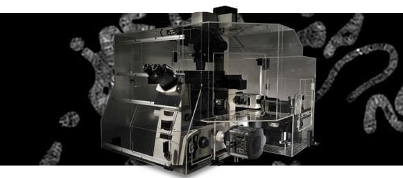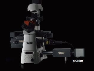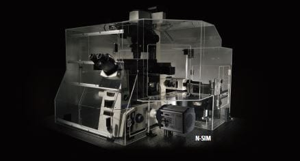


Structured illumination super-resolution microscope delivering twice the resolution of traditional diffraction limited microscopes.
Super-resolution microscope visualizing cellular structures and molecular activity at resolutions never before achieved by conventional light microscopy.
Nikon’s N-SIM microscopy system can produce twice the resolution of conventional optical microscopes. Using an innovative approach based on Structured Illumination Microscopy technology licensed from UCSF, the N-SIM enables detailed visualization of minute intracellular structures and their functions.
N-SIM doubles the resolution of conventional optical microscopes (~115nm in 3D SIM mode*) by combining “Structured Illumination Microscopy” technology licensed from UCSF with Nikon’s renowned CFI Apo TIRF 100x oil objective lens (N.A. 1.49).
* resolution will depend on laser wavelength and imaging mode.
The SR (super-resolution) objectives have been designed for new applications that break the diffraction barrier.
The most recent optical designs using wavefront aberration measurement have been applied to yield superb optical performances with the lowest asymmetric aberration.

N-SIM provides the fastest imaging capability in the industry, with a time resolution of 0.6 sec/frame, effective for live-cell imaging.


The 3D-SIM illumination technique improves axial resolution to 269 nm and has the capability of optical sectioning of specimens, enabling the visualization of more detailed cell structures at higher spacial resolutions.

This mode captures super-resolution 2D images at high speed with incredible contrast. TIRF-SIM mode takes advantage of Total Internal Reflection Fluorescence observation at double the resolution as compared to conventional TIRF microscopes, facilitating a greater understanding of molecular interactions at the cell surface.
|
|
|

By attaching two EMCCD cameras to the microscope with the optional Two Camera Imaging Adapter*, simultaneous two-wavelength super-resolution imaging with excitation of 488 nm and 561 nm is possible.
* Andor Technology Ltd.

Two Camera Imaging Adapter (for N-SIM)
* The actual product may differ slightly in design.
| Super-Resolution | Lateral (XY): ~85-110nm (dependant on wavelength and optics)* Axial (Z): ~300nm (dependant on wavelength and optics)* 3D Axial Range: up to 20μm |
|---|---|
| Image acquisition time | Up to 0.6 sec/frame (TIRF-SIM/2D-SIM)Up to 1 sec/frame (Slice 3D-SIM)
(needs more 1-2 sec. for calculation) |
| Imaging mode | TIRF-SIM2D-SIM
Slice 3D-SIM Stack 3D-SIM |
| Multi-color imaging | Up to 5 colors |
| Compatible Laser | Standard: 488nm, 561nmOption: 405nm, 458nm, 514nm, 532nm, 640nm
Laser combination: 405 nm/488 nm/514 nm/532 nm/561 nm, 405 nm/488 nm/514 nm/561 nm/640 nm, 458 nm/488 nm/514 nm/532 nm/561 nm, 458 nm/488 nm/514 nm/561 nm/640 nm |
| Compatible microscope | Motorized inverted microscope ECLIPSE Ti-EPerfect Focus System
Motorized XY stage with encoders Piezo Z stage |
| Compatible objective | CFI SR Apochromat TIRF 100×oil (NA1.49)CFI Apochromat TIRF 100×oil (NA1.49)
CFI SR Plan Apochromat IR 60×WI (NA1.27) CFI Plan Apochromat IR 60×WI (NA1.27) |
| Camera | EM CCD camera iXon3 DU-897E (Andor Technology Ltd.) |
| Software | NIS-Elements Ar/NIS-Elements C (for Confocal Microscope A1+/A1R+)Both require optional module software NIS-A N-SIM Analysis |
| Operating conditions | 20°C to 28°C ( ± 0.5 oC) |
*Resolution depends on dye properties
 Bioengineering
 Biomechanics
 Cardiovascular Research
 Cell Biology
 Dermatology
 Developmental Biology/Embryology
 Hepatology
 Microbiology
 Molecular Biology
 Nephrology
 Neurobiology / Neuroscience
 Obstetrics & Gynaecology
 Oncology
 Ophthalmology
 Rheumatology
 Stem Cell & Regenerative Medicine
 Tropical Medicine
 Urology
 Vascular Research