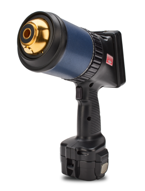
Emittance at 3-5 and 8-12 microns
The ET-10 Emissometer answers the long-standing need for measuring emissivity as an entry parameter for calibrating infrared thermography devices. The ET-10 measures total reflectance and directional emissivity at 20 degrees angle of incidence for 3-5 and 8-12 microns. The 8-12 band can optionally be extended to 14 microns. View results immediately on the touchscreen display.
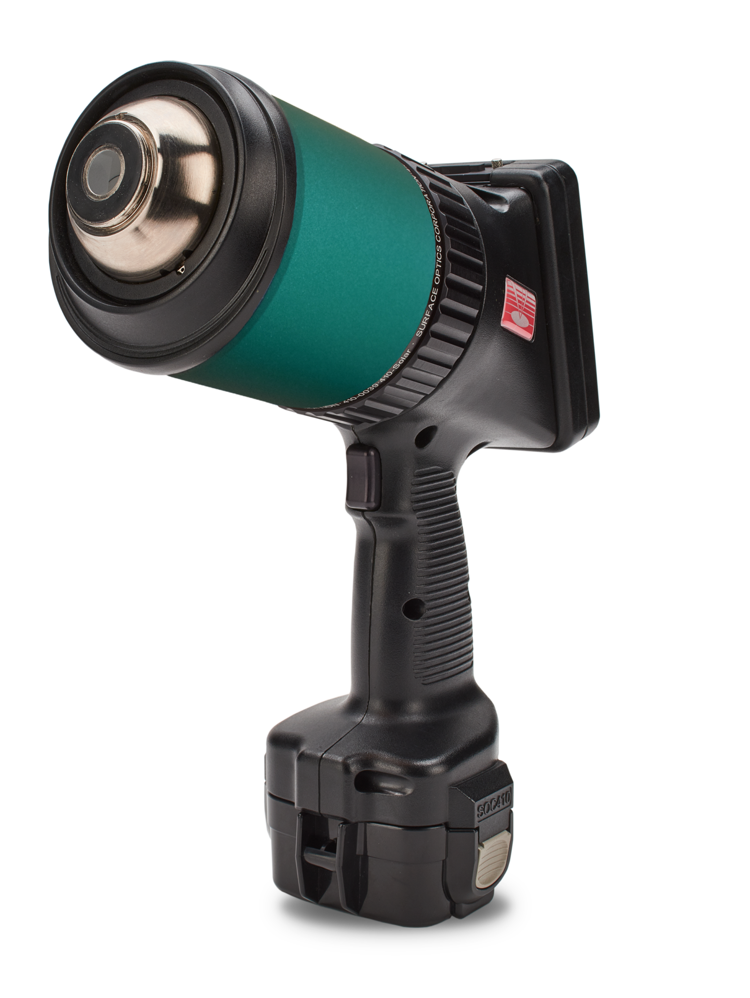
Reflectance at 850nm, 905nm, 940nm and 1550nm
The 410-LIDAR measures reflectance simultaneously at four key wavelengths employed by lidar systems. Collect measurements on materials and objects that cannot easily be brought into the lab. Fast calibration and measurement times without sacrificing measurement accuracy and repeatability. Option to customize wavelengths.
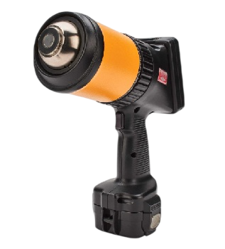
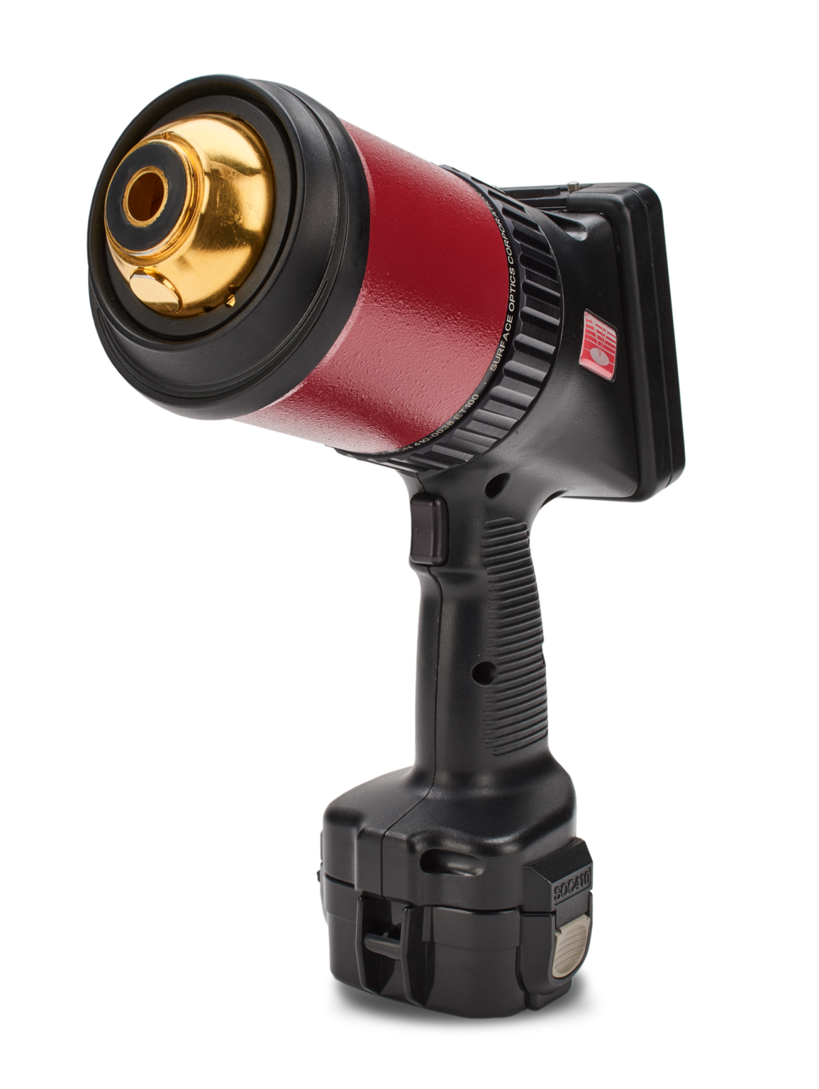
Infrared reflectance measurements
The 410-DHR is a product of collaboration between U.S. Naval Air Systems Command and Surface Optics, with vital input from the Naval Research Lab (NRL) and National Institute of Standards and Technology (NIST). It was developed to answer a broad need for a portable device for the verification of the optical properties of large objects in the field. The 410-DHR measures the integrated surface reflectance of a surface at two angles of incidence (20° and 60°) and for six discreet wavelength bands in the .9 to 12 μm spectral range.
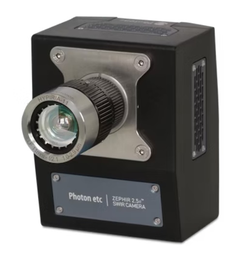
The ZephIR 2.5e is an affordable deep-cooled, SWIR camera, sensitive from 1000 to 2500 nm.
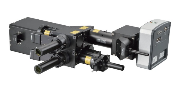
Modular illumination system with ultimate flexibility and expandability.
The Nikon Ti2-LAPP system provides modular illuminators for total internal reflection fluorescence (TIRF), photoactivation/conversion, photobleaching, epi-fluorescence, as well as super-resolution microscopy (N-STORM). Each module can be flexibly combined to build microscope systems that are optimized for individual research needs. For example, multiple TIRF modules can be incorporated into a single microscope for anisotropy experiments and fast, multi-angle TIRF imaging. Combined with the Ti2’s stratum structure, up to five illumination modules can be incorporated into a single microscope (e.g. two TIRFs, a FRAP, a DMD, and an Epi-FL module can all be integrated into one Ti2).