Microscope Camera Head DS-Fi1c

5-megapixel cooled color digital microscope camera head ideal for acquiring detailed images under a variety of illumination techniques.

A 5-megapixel cooled color digital microscope camera head ideal for both brightfield and fluorescence applications.
Featuring a high definition cooled color camera head and the latest optical technology, the DS-Fi1c produces stunning high-resolution images of up to 2560 x 1920 pixels. Its Peltier cooling mechanism, high dynamic range, low noise and high frame rates achieves high performance under short and longer exposure times, making it suitable for sensitive samples across a wide variety of applications including fluorescence,brightfield, phase contrast and differential interference contrast (DIC).

Microscope Camera Head DS-Vi1 High Speed Microscope Camera

High speed, high sensitivity, microscope camera featuring 2-megapixel color CCD with outstanding SXGA video display rates of up to 27 frames per second.

A digital microscope camera head offering high speed, high sensitivity and high dynamic range.

Nikon has added the high speed DS-Vi1 color microscope camera to its market-leading range of Digital Sight cameras for microscope imaging. The DS-Vi1 high speed color camera head offers high frame rates and increased sensitivity for both multiple live image and sharp still image capture, making it ideal for high end biological research, clinical analysis and documentation as well as medical teaching use.

PicoQuant Time-resovled Confocal Fluorescence Microscope – MT200

Time-resolved Confocal Fluorescence Microscope with Unique Single Molecule Sensitivity

Picoquant Time-resolved Confocal Fluorescence Microscope – MicroTime 200

  • Complete confocal system with laser combining unit, inverted microscope body and multichannel detection unit
  • Turn-key diode lasers for multicolor excitation from 375 to 900 nm
  • Up to 4 truly parallel detection channels using application-optimized detection with SPADs, PMTs or Hybrid-PMTs
  • Time-Correlated-Single Photon Counting (TCSPC) and TTTR mode for investigation of fast dynamics in FCS and FLIM
  • Piezo scanning for 2D- and 3D-lifetime imaging and accurate point positioning
  • Two optional exit ports for additional hardware, e.g. spectrographs
  • Advanced easy-to-use data acquisition, analysis and visualisation software SymPhoTime 64
  • Unique upgrades to 2focus FCS, simultaneous AFM/FLIM, UV measurements
  • STED add-on for super-resolution imaging 

TechnoSpex – uRaman-Modules

The uRaman-Module can be easily integrated in most major brands upright microscope (Nikon, Olympus, Leica, and Carl Zeiss).

The uRaman is the state-of-the-art flexible Raman spectroscopy system. It offers high performance, ease of use and yet remains cost effective for tight budget users.
The uRaman-Module can be easily integrated with most upright microscopes from the following brands: Nikon, Olympus, Leica, Carl Zeiss, etc. It can also possible to integrate with existing imaging setup, such as fluorescence microscopy, without affecting overall microscopy performance. Multiple modules, stackable to achieve dual or triple wavelength measurement.

Integrated with a 532nm, 633nm or 785nm laser and a high sensitivity linear array detector, the module includes power attenuators, laser safety interlock and user friendly software.
Check with our Application Scientist for compability of your microscope.

TechnoSpex – uRaman-Ci Raman Micro-Spectroscopy System

The uRaman Ci is a complete Raman Micro-Spectroscopy System that comes with the Nikon Ci-L research grade upright microscope.

The uRaman-Ci is a complete Raman Micro-Spectroscopy System that consists of the uRaman module being integrated with the Nikon Ci-L research grade upright microscope.
In addition to Raman and bright field microscopy, the uRaman-Ci can also be equipped with other multimodality imaging capabilities such as reflected bright field, dark field, DIC, polarization and fluorescence imaging. We also have a full range of Nikon Objective Lens and accessories such as cuvette holder for measuring liquid samples for you to choose from. uRaman-Ci can also be equipped with a XY motorized stage and uSoft Map software for Raman chemical mapping.

uRaman-Ci can also be equipped with a XY motorized stage and uSoft Map software for Raman chemical mapping.

Request a quote to know Raman microscope price.

Cytoviva Hyperspectral Microscopy

Cytoviva Hyperspectral Microscopy – Visible Near Infrared (VNIR) and Short Wave Infrared (SWIR)

CytoViva’s Hyperspectral Imaging technology was specifically designed to provide quantitative spectral analysis of nanoscale materials imaged with the patented CytoViva darkfield-based microscope system or with other microscopy modalities

This can include spectral analysis of both biological and materials-based nanoscale samples, which may be isolated or integrated in cells, tissue or other materials-based matrices. CytoViva provides hyperspectral imaging in the visible near-infrared (VNIR 400nm-1,000nm) wavelength ranges as well as in the short wave infrared (SWIR 900nm-1,700nm) wavelength ranges.

In addition, CytoViva provides hyperspectral imaging in both wavelength ranges for macro-level samples that do not require microscopy level analysis.


Hyperspectral images appear very similar to a traditional optical image with one important difference. When observed via image analysis software, each pixel of a hyperspectral image provides the complete spectral response of that pixel’s spatial area within the VNIR or SWIR spectral range. At 100x magnification, a hyperspectral image may contain as many as 700,000 pixels as small as 128nm each. This spectral data is recorded at approximately 2nm of spectral resolution in the VNIR range, enabling minute spectral differences to be measured from pixel to pixel within the image.

Using CytoViva’s proprietary hyperspectral image analysis software, it is possible to identify, locate and map nano-scale materials within a sample based on the unique spectral response of this material. This can be accomplished without any special staining or tagging of the target material, as is often required with traditional microscopy techniques. In addition, when functional groups are added onto or into a nano-scale sample, they can often be identified, as this change in the sample’s surface chemistry will be spectrally detected.

The primary components of CytoViva’s Hyperspectral Imaging technology include a transmission diffraction grating spectrograph and an integrated camera. These components are mounted onto the camera (C) mount of the microscope. A hyperspectral image is created in a line scan or “pushbroom” fashion, by moving the sample across the field of view of the microscope and spectrograph via an automated translational microscope stage. Typically these spectral images are created in seconds or minutes, depending on the required exposure. The hyperspectral imaging hardware is integrated with customized image analysis software. This proprietary software provides the ability to compare spectra within a sample image or between images. It can also build a spectral library of unique samples elements. Using this spectral library, these sample elements can then be mapped in subsequent samples.

To learn more about the research applications supported by CytoViva’s Hyperspectral Microscopy System, please click on the hyperspectral applications page above.

Cytoviva Enhanced Darkfield Microscopy

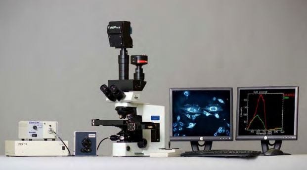
The CytoViva® Microscope System – Enhanced Darkfield Microscopy


Cytoviva Enhanced Darkfield Microscopy – Specifically designed to support research in nanotechnology and infectious disease, CytoViva employs a patented (US patents No. 7,542,203, 7,564,623) enhanced darkfield-based optical illumination system.

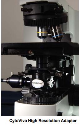
This structured illumination technology replaces the standard condenser on a research grade microscope. By improving the alignment and focus of darkfield or oblique angle illumination, the technology enhances signal-to-noise of nanoscale samples up to seven times over standard darkfield optics. This enables scientists to optically observe a wide range of nanoscale materials quickly and easily in solution, live cells, tissue and materials based matrices. In addition, non-fluorescent live cells and pathogens can be easily observed at a level of detail not possible with traditional optical imaging techniques such as phase contrast or differential interference contrast.

When using the CytoViva Dual Mode Fluorescence system, researchers can also observe the interactions between fluorescently labeled nano-particles or bacteria and live unlabeled cells. This unique capability can eliminate the need to create computer enhanced overlay images which require two different illumination methods and advanced software programs. Finally, when combined with CytoViva’s Hyperspectral Imaging capability this high signal-to-noise microscopy method enables researchers to spectrally characterize and map nanoscale samples in a wide range of environments.


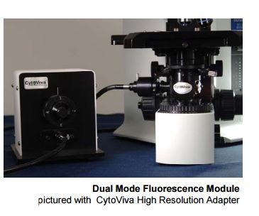
Dual Mode Fluorescence Module

cytoviva dual fluorescence module

The Dual Mode Fluorescence (DMF) module allows for the observation of both fluorescent and non-fluorescent sample portions simultaneously and in real-time. Samples are viewed directly through the microscope eyepiece and captured using a standard microscope camera without the need for complicated software or electronic manipulation.

The Environmental Chamber

Live Cell Chamber

The CytoViva environmental chamber supports high resolution, long-term studies of live cells, while enabling real-time, simultaneous observation of fluorescent and non-fluorescent sample portions.

Manufactured by Warner Instruments, the CytoViva environmental chamber is a modified version of the Warner RC-30 Confocal chamber. This modification supports oil immersion contact with the CytoViva high resolution illumination system as well as an oil immersion microscope objective.

The CytoViva system supports all traditional environmental chamber applications including perfusion, temperature control and gasses. Operating as a closed bath system, the CytoViva compatible chamber works with both inverted and upright research grade microscopes.

Now researchers can observe, track and image the interactions between live cells and other elements such as fluorescently labeled nano-particles and bacteria. These observations can be made over hours or even days.

Traditional cell biology and neuroscience applications are also supported by this system.
With the CytoViva system and environmental chamber you can observe fluorescently labeled cellular components simultaneously with the unlabeled portions of the cell.

The CytoViva Environmental Chamber can also be utilized as a fully functional micro-fluidics platform supporting applications in drug delivery, tissue engineering and layer assembly of nano-materials.

Cytoviva Enhanced Darkfield Illumination System

The CytoViva® Microscope System – Advanced Darkfield Illumination System

Advanced Darkfield Illumination System


The CytoViva advanced darkfield illumination system replaces the standard microscope condenser. The specialized illuminator focuses fixed-geometry, highly collimated light at oblique angles on the sample. This serves to dramatically improve contrast and signal-to-noise ratio which allows for optimized resolving power and detection capability of non-fluorescing samples.

cytoviva high resolution adaptor

Cytoviva 3D Enhanced Darkfield Imaging System

The CytoViva 3D Enhanced Darkfield Imaging System

The CytoViva 3D Enhanced Darkfield Imaging System provides a method for locating non-labeled nanostructures (particles, tubes, etc.) in a variety of translucent matrices (cells, tissue, organisms). This technique leverages the high signal-to-noise optical performance of the patented CytoViva Enhanced Darkfield Microscope Technology in combination with patent-pending deconvolution and particle location routines to provide users with a three dimensional optical model of their sample. Most importantly, this technique does not require the use of fluorescent labels on the nanoparticles to obtain an image of the particles, thus removing the potentially negative influence of these labels on the sample.

homer-head-and-cnts-1024x699.jpg

The 3D image is created by acquiring and storing a “stack” of equally spaced two dimensional images using the CytoViva Enhanced Darkfield Microscope Technology, a piezo-driven Z-axis stage and CytoViva’s proprietary image acquisition and control software.  This stack of images is then processed using CytoViva 3D Image Analysis ImageJ plug-ins to locate the subject particles, as well as, deconvolve the surrounding cells, tissue or other translucent matrix.  The resulting optical image is then viewed in an ImageJ 3D viewer.  The use of ImageJ also allows further processing of the data in an environment familiar to many users.

The images above  illustrate three examples of  the 3D capability.  Figure 1 is a 3D deconvolution  of nanoparticles  (NPs) in a cell with the NPs in red, the nuclear structure in blue and the  cell structure in gray. Figure 2  is a 3D deconvolution of a carbon nanotube piercing a tissue section.

The CytoViva 3D Enhanced Darkfield Imaging System is designed to facilitate research in a variety of applications in nanobiology (targeted drug delivery and pathogen detection), nanotoxicology (nanoparticles and carbon nanotubes in tissue), and others where it is useful to understand the location of nanostructures in a three dimensional space.  This capability is fully compatible with CytoViva’s Hyperspectral Imaging system and can be operated on the same microscope platform, thus allowing existing systems to be easily upgraded to include this capability.

3D Videos:

Unlabeled Au Nanoparticles in a Cell

Unlabeled Carbon Nanotubes Embedded in Tissue

Fiber Matrix

PicoQuant Fluorescence Lifetime Spectrometer – FluoTime 250

PicoQuant Fluorescence Lifetime Spectrometer – FluoTime 250 integrates all essential optics and electronics for time-resolved luminescence spectroscopy in a compact, fully automated device.

Compact and Modular Fluorescence Lifetime Spectrometer

Fully automated compact and modular system

  • Filter based emission selection, optional monochromator
  • Supports both TCSPC and Multi-Channel Scaling data acquisition
  • Step by step application wizards and scripting option
  • Covers lifetime ranges from picoseconds to milliseconds

A compact spectrometer for routine and complex measurements

PicoQuant Fluorescence Lifetime Spectrometer – FluoTime 250 integrates all essential optics and electronics for time-resolved luminescence spectroscopy in a compact, fully automated device. This spectrometer is designed to assist the user in carrying out routine as well as complex measurements easily and with high reliability thanks to fully automated hardware components and a versatile system software featuring wizards providing step-by-step guidance.

Fully automated with high performance optics

FluoTime 250 - Fully automated lifetime spectrometer with high performance optics

All components (attenuators, polarizers and filter wheel) in the optical beam paths are motorized and controlled by the EasyTau 2 software. Emission wavelength selection is implemented via a motorized filter wheel equipped with various cut-off or bandpass filters as selected by the customer. An optional monochromator is available for the UV/VIS spectral range.

Flexible excitation sources

PicoQuant's PDL 820 laser dirver with LDH laser head and pulsed diodes from the PLS Series

The FluoTime 250 uses picosecond pulsed lasers diodes and LEDs from PicoQuant’s LDH and PLS series, covering a spectral range from 255 to 1550 nm. All excitation sources are controlled via a PDL 820 laser driver, which allows not only to vary output power and repetition rates (up to 80 MHz) but also supports burst mode for selected laser heads.

TCSPC and MCS based data acquisition

TimeHarp 260 - TCSPC and MCS board with PCIe interface

Several outstanding data acquisition units are available with various temporal resolutions (from 4 ps to 250 ns). Coupled with the right detectors from the PMA or PMA Hybrid Series, the FluoTime 250 is capable of measuring time-resolved emissions with lifetimes ranging from approximately 10 ps up to several hundreds ms.

A choice of detectors

Hybrid photomultiplier detector assembly from the PMA Hybrid Series

The FluoTime 250 can be equipped with a single photon counting detector from either the PMA or PMA Hybrid Series.These detectors offer picosecond temporal resolutions and cover different spectral ranges between 180 and 920 nm. For studies in the near infrared, a PMT module with sensitivity from 950 to 1400 nm is also available. Each detector includes an electro-mechanical shutter, active cooling, and overload protection.

System software EasyTau 2

Screen shot the EasyTau 2 software analyzing the fluorescence decay of antracence

The EasyTau 2 software package is the one-stop solution for both full hardware control of the FluoTime 250 as well as for interactive data analysis and fitting. The software offers a Windows-based graphical user interface with dedicated applications wizards guiding users through the optimization and data acquisition process for many common applications. Advanced users get full control over all instrumental aspects thanks to a customized mode and scripting language. The integrated analysis and fitting module supports a broad range of time-resolved spectroscopy applications, such as fluorescence and phosphorescence decays, or anisotropy measurements. A powerful report generator allows creating presentation-ready graphical and numerical output.