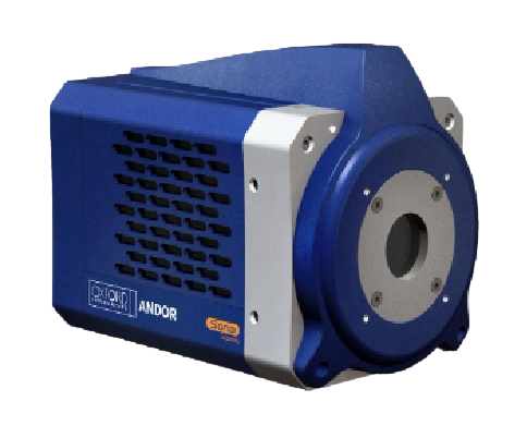
Sona-11: For the Widest Field of View
Sona-11 has the biggest sensor on the market with a full 32 mm sensor diagonal! Andor’s unique technology usefully accesses the entire 2048 x 2048 array offering 62% larger field of view than competing back illuminated cameras. The 95% QE and larger pixel size of 11 µm provides optimal photon collection, for the most light-starved applications. Study structures and processes within the cell in perfect resolution using techniques such as confocal, TIRF and Single Molecule Localization Microscopy (SMLM)
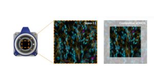
See the full picture: With a 32 mm sensor diagonal Sona-11 has a field of view advantage:
✔️2.9x larger field of view vs typical sCMOS
✔️2.1x larger field of view vs 22 mm format sCMOS
✔️62% larger field of view vs competing back-illuminated sCMOS (1608×1608 array)
✔️Capture weak signals – 95% QE is complemented by large 11 µm pixel
size for optimal photon collection
✔️SRRF-Stream+ Super-Resolution – Transform a standard microscope to super-resolution!
✔️NEW Python ready – Updated camera SDK integrates a Python
wrapper for speedy integration
Key Applications:
- Developmental biology
- Neuroimaging
- Super-resolution
- Transcriptomics
- Intracellular trafficking
- Plasma membrane studies
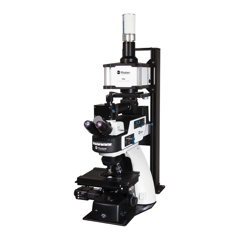
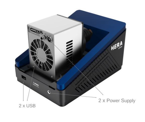
Global hyperspectral microscope
IMA is a hyperspectral microscope delivering spectral and spatial information in the VIS, NIR, and SWIR range (400 nm – 1620 nm). This system rapidly maps photoluminescence, electroluminescence, fluorescence, reflectance, and transmittance. Based on high throughput global-imaging filters, IMA is faster and more efficient than standard point-by-point or line-scan based systems.
IMA Applications Overview
- Perform complex material analyses such as solar cell characterization and semiconductor quality control
- Study of sample formation, degradation and identification of deficient areas
- Imaging of multiplexed emitters
- Access to the second biological window (900 – 1620 nm)
- Mapping of spectral heterogeneities
- Study IR markers in complex environments including live cells and tissue.
- Fast imaging – 1.4 million spectra in minutes
- Retrieve darkfield images and obtain a contrast of transparent and unstained samples such as polymers, crystals or live cells.
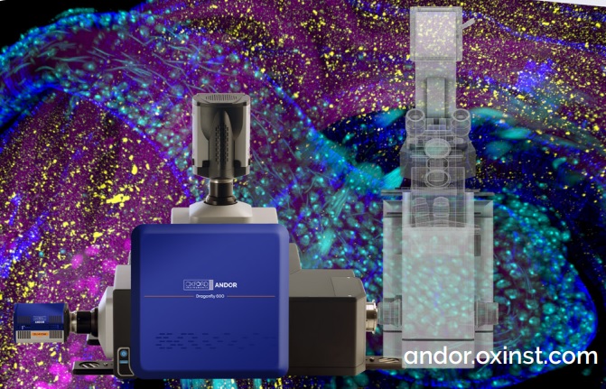
Dragonfly High Speed Confocal Microscope System
Dragonfly delivers outstanding multi-dimensional images from subcellular (nm) to whole organism (cm), while significantly boosting productivity.
Its unrivalled combination of speed and sensitivity allows researchers to discover unforeseen dynamic events and image live organisms for days. The new B-TIRF and super resolution modules reveal smaller details, such as the dynamics of viral infection and the ultrastructure of chromatin or organelles.
Dragonfly is a high-contrast multi-dimensional imaging platform capable of four key imaging modalities. At its core is a multi-point confocal for high-speed and high-sensitivity imaging. Capturing at speeds at least 10x faster than conventional confocal technology, with dramatically improved sensitivity from 400-800 nm. Dragonfly is the optimal solution for live cell imaging, providing low phototoxicity and photobleaching, or perfect for fast volume acquisition of fixed samples.
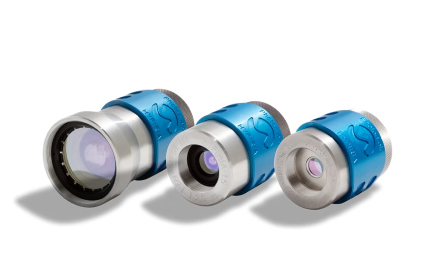
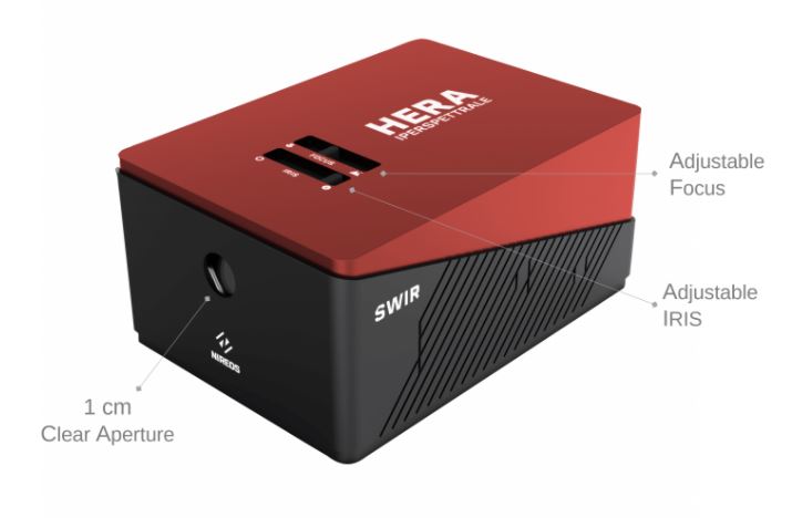
SWIR Lenses – 1.0 to 2.9 µm
The HypIRia are wide-aperture, image-space telecentric lenses precisely crafted to provide clear and bright images in the SWIR spectral range. A broadband anti-reflective coating on carefully selected glass delivers high throughput and low-aberration imaging on a range extending from 1.0 to 2.9 µm.
A translational focus provides excellent stability during focusing. Robustly built, they are well suited for industrial applications or field work.
Three focal lengths are currently available.
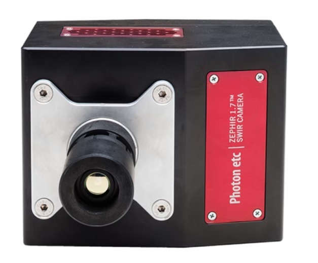
Deeply Cooled InGaAs Camera
The ZephIR 1.7 is a high-end, scientific grade, 640 x 512 pixels resolution, InGaAs camera that marries performance with reliability. It has extremely low noise levels, high efficiency, and a rapid frame rate compatible with an external trigger. This is made possible by a combination of state-of-the-art control electronics and a four stage thermoelectric cooler (TEC) which can maintain an operating temperature as low as -80 °C. The TEC, in turn, uses forced air cooling which requires none of the maintenance of a water or liquid nitrogen chilled unit.
The ZephIR 1.7 is one of the most sensitive and dependable InGaAs cameras on the market.
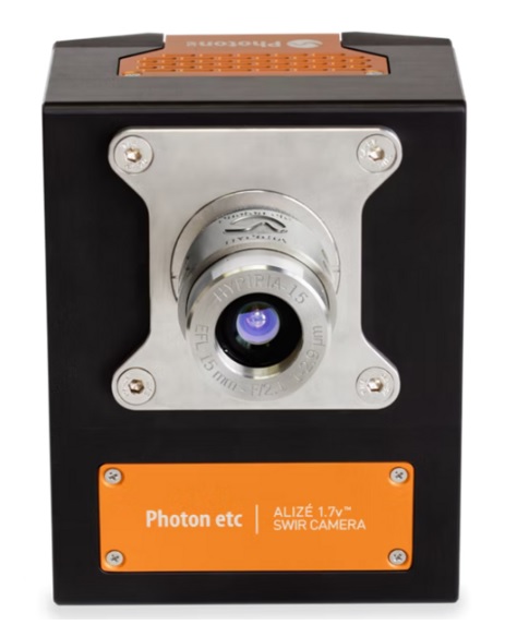
The Alizé 1.7 is a high-end, scientific grade, 640 x 512 pixels resolution, InGaAs camera that marries performance with reliability.
NIR imaging with this versatile InGaAs camera
The Alizé 1.7 is a high-end, scientific grade, 640 x 512 pixels resolution, InGaAs camera that marries performance with reliability. It has low noise levels, high efficiency, and a rapid frame rate compatible with an external trigger. This is made possible by a combination of state-of-the-art control electronics and a four-stage thermoelectric cooler (TEC) which can maintain an operating temperature as low as -50 °C. The TEC, in turn, uses forced air cooling which requires none of the maintenance of a water or liquid nitrogen chilled unit.
The Alizé 1.7 is amongst the most cost-effective high-end InGaAs cameras on the market.
Key Features
- Compact
- Highly reliable
- Long lifetime
- No maintenance
- Low dark current
- Low readout noise
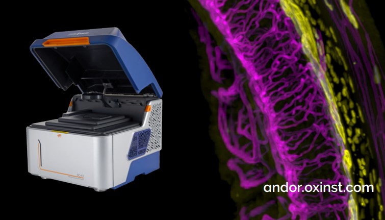
Andor’s new Benchtop Confocal delivers amazing images at the push of a button. This plug and play laser confocal system has been designed with cost, performance and accessibility in mind, ensuring hassle-free, high-quality 2D & 3D imaging for busy researchers.
*Microscopy Today 2022 Innovation Award Winner*
Fast and Easy Confocal Microscopy in any Lab
BC43 is a super-compact unit that is rich in features and benefits, making it the ideal microscope for early-stage researchers and experienced microscopists alike. With no requirement for a darkroom, researchers can access laser confocal microscopy technology on the same bench as their samples. This, coupled with the fast learning curve, allows individual users and entire labs to become much more productive, without compromising results.
Powerful and Intuitive Acquisition Software
At the heart of BC43 is a software that is not only feature-rich and flexible to your needs, it is easy-to-use as well:
- Imaris rendering engine – stunning real-time 3D images
- Clear and concise user-interface – acquire images in no time
- Multi-dimensional imaging protocols – advanced 3D time lapse or multi-position montage made easy