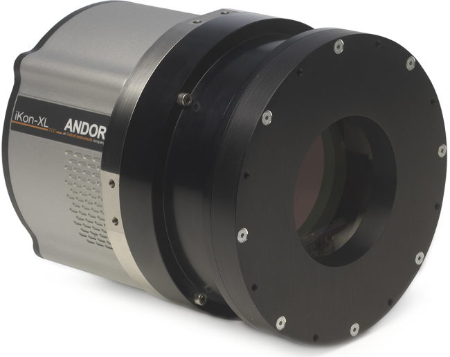
Andor’s iKon-XL is a thermoelectrically (TE) cooled, very large area CCD camera platform, accommodating large field of view sensors that are ideally suited to long exposure astronomy applications. Using the unique, patent-pending ColdSpace™ technology, the ultrasensitive iKon-XL is capable of TE cooling a 16.8 Megapixel sensor (e2v back-illuminated) down to -100 °C, avoiding the requirement for liquid nitrogen or unreliable cryo coolers. The iKon-XL also uses an exclusive Extended Dynamic Range technology, facilitating lowest noise and maximum well depth within one scan, complemented by up to 18-bit digitization capability. Flexible connectivity is standard through either USB 3.0 or a long distance direct fibre optic interface.
Andor’s PV Inspector NIR Camera is designed to offer ultimate speed and sensitivity performance for in-line Electro- and Photoluminescence Inspection, delivering > 90% QE beyond 800 nm and incorporating Fringe Suppression Technology™ to minimize fringing effects in the NIR
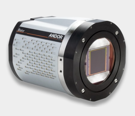
Balor is Andor’s game-changing, very large area sCMOS camera platform for Astronomy. It launches with the Balor 17-12 model, featuring an enhanced FoV 16.9 Megapixel, 70mm diagonal sensor, coupled with fast, low noise readout capability. Balor is ideal for measuring photometric and astrometric variability from milliseconds to tens of seconds timescales.
Balor is the largest commercially available sCMOS camera, designed for ‘dynamic astronomy’ applications such as Orbital Debris tracking, Solar Astronomy, Solar System Object detection, Exoplanet Discovery, Atmospheric Studies and Fast Time Resolution Astrophysics. Balor lends itself particularly well to the ‘atmospheric freezing’ techniques of Speckle/Lucky Imaging, enabling resolution enhancement of ground-based astronomy over a much larger field of view than is readily achievable through use of adaptive optics.
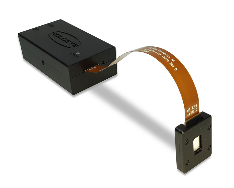
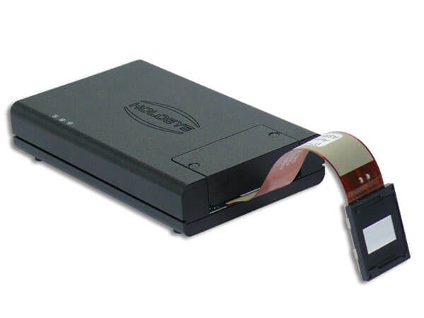
The LUNA phase only Spatial Light Modulator (SLM) consists of a driver unit with standard digital video interface (DisplayPort) and a phase only LCOS (Liquid Crystal on Silicon) microdisplay with full HD resolution (1920 x 1080 pixel) and 4.5 µm pixel pitch leading to an active area diagonal of 0.39” with an aspect ratio of 16:9.
The LUNA Spatial Light Modulator is our most compact SLM platform for integration into small sized or even portable solutions.
The LUNA phase only Spatial Light Modulator (SLM) consists of a driver unit with standard digital video interface (DisplayPort) and a phase only LCOS (Liquid Crystal on Silicon) microdisplay with full HD resolution (1920 x 1080 pixel) and 4.5 µm pixel pitch leading to an active area diagonal of 0.39” with an aspect ratio of 16:9.
The driver ASIC is embedded in the LCOS microdisplay itself. This saves board space which enables a very compact driver and makes integration more convenient. The standard driver box has a size of only 84.4 x 47 x 28.8 mm. The display can even accept video data input via a 4-lane MIPI DSI. This brings phase only Spatial Light Modulator technology to a new level of potential for industrial implementations.
The LUNA SLM usees fast full digital addressing which assures high reliability and a compact driver unit. A certain gray level represents a defined average voltage across the LC cell. This voltage leads to a variable tilt of the LC molecules due to their electrical anisotropy. As LC molecules also show optical anisotropy this tilt changes the refractive index of the LC molecules (for incident polarization along the long display axis) which causes a modified optical path length within the LC cell. The addressed gray level is converted into a phase level.
The LUNA driver unit is equipped with an USB interface for changing the voltage vs. phase level distribution (gamma control) and dynamic range (voltage across the LC cell) in order to calibrate the SLM for different wavelengths.
The LUNA SLM is a plug & play phase modulator and can be addressed with phase functions via standard graphics cards as extended monitor device. Addressing can be done using the supplied Pattern Generator software, the HOLOEYE Slideshow Player software or standard image viewer software. HOLOEYE also provides an SLM Display Software Development Kit (SDK) which provides APIs (Application Programming Interface) for different programming languages.
LUNA Spatial Light Modulator – Microdisplay Features
Display Type:
Reflective LCOS (Phase Only)
Resolution:
1920 x 1080
Pixel Pitch:
4.5 µm
Fill Factor:
91 %
Active Area
8.64 x 4.86 mm (0.39″ Diagonal)
Addressing
8 Bit (256 Phase Levels)
Signal Formats
DisplayPort – HDTV Resolution
Input Frame Rate
60 Hz
LUNA Phase Only Spatial Light Modulator Versions
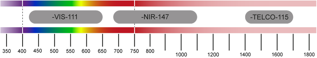
HOLOEYE offers different phase display versions which can be driven with the LUNA driver unit. The different display versions are optimized for the use at different wavelength ranges. Currently we offer a VIS version (420 – 650 nm) and a version for the telecommunication waveband around 1550 nm (1400-1700 nm).
Version
Wavelength Range
Reflectivity
Max. Phase Shift
LUNA-VIS-111
420 – 650 nm
61-67%
4.3 π @ 450 nm
3.2 π @ 520 nm
2.4 π @ 635 nm
LUNA-TELCO-112
1400 – 1700 nm
70 %
2.3 π @ 1550 nm
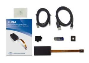
LUNA Spatial Light Modulator Kit – Contents:
LUNA Spatial Light Modulator – Scope of Supply
LUNA phase display (1920 x 1080 pixel) incl. flex cable
LUNA driver unit
USB Cable
DisplayPort / Mini DisplayPort cable
Magnetic display mount & display adaptor
Screw adaptor M4 to ½”
Safety instructions & quick-start guide
USB flash drive with documentations, detailed manual, Configuration Manager and SLM software
LUNA Phase Modulator – Software Features:
The LUNA phase only spatial light modulator devices can simply be addressed like an external monitor using the standard DisplayPort interface of the graphics card. No additional software or dedicated hardware is needed to operate the SLM.
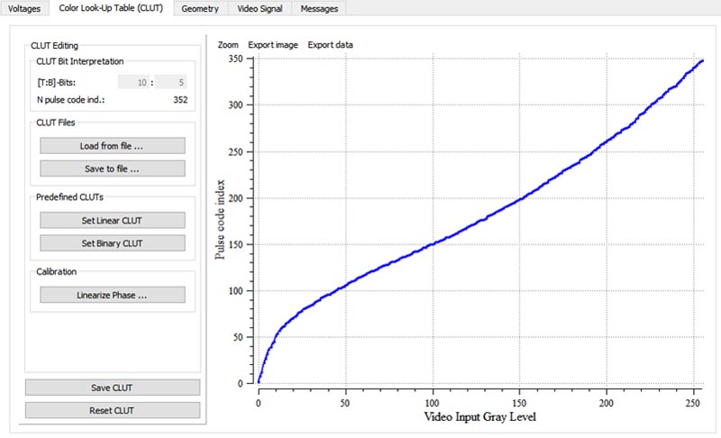
The device is delivered with a Configuration Manager software. The Configuration Manager can be used to change geometrical settings, brightness, contrast and the electrooptical response by applying a new gamma curve or another digital drive scheme. For these advanced calibrations the device uses an USB connection to a virtual COM port of the computer.
Computation of computer generated holograms (CGH) from user defined images
generation of SLM signals representing basic optical functions such as lenses, gratings, axicon and vortex functions
superposition of CGH’s with basic optical functions to combine functionalities
Besides that a special SLM Slideshow Player Software comes with the device. Also an SLM Display SDK is available for download which provides APIs (Application Programming Interface) for different programming languages to show images and data/ phase arrays directly on a HOLOEYE Spatial Light Modulator:
National Instruments LabVIEW™ 8.6 and later
MathWorks MATLAB® R2009b and later
Octave 5.2
Python™ 2.7 and 3.x
C or C++ compiler (e. g. Microsoft Visual C++ Compiler)
Display Dimensions (Unit: mm):
The current LUNA microdisplay package has a size of only 25.7 x 12.6 x 3.4 mm. For more convienient mounting we deliver it with a display mounting adaptor.
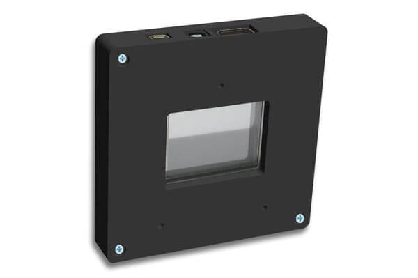
The LETO-3 phase only Spatial Light Modulator is based on a reflective LCOS microdisplay with full HD (1920 x 1080 pixel ) resolution. With a pixel pitch of 6.4 µm and a small interpixel gap of 0.2 µm the LETO-3 SLM provides a high fill factor of 93%.
The LETO-3 Spatial Light Modulator offers a reflectivity between 62% up to 75% (dependent on wavelength and version). The device offers diffraction efficiencies of more than 80% (16 level blazed grating) which leads to a total light efficiency of ~ 60%.
The LETO-3 SLM driver is prepared to work in color-field-sequential (CFS) mode e.g. with color-switchable LASER or LED lighting and the device features an LED-connector which can be used to synchronize the light source with the device. The driver features 4 slots with preconfigured CFS configurations for different environments which can be slected using the Configuration Manager Software.
Versions
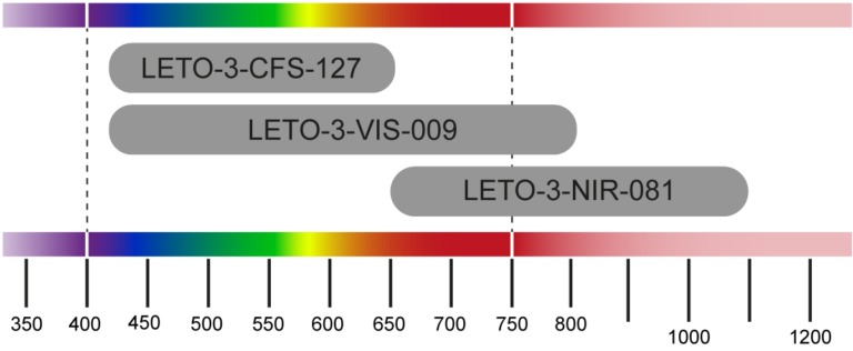
HOLOEYE offers three different LETO-3 Spatial Light Modulator versions which are optimized for the use at different wavelength ranges / for different applications. One SLM version is optimized for the visible range, one version for the near IR range up to 1100 nm and a third version is optimized for fast response (180 Hz) and use for color-field-sequential (CFS) operation in the visible range with color-switchable RGB Laser.
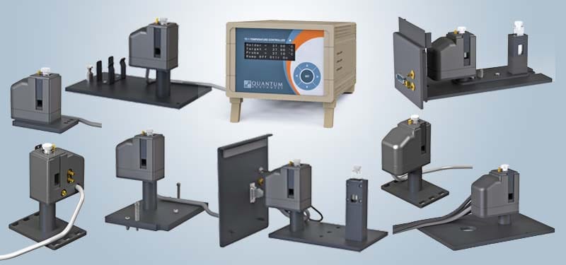
The t2 Sport™ is a Peltier-Temperature controlled cuvette holder with two optical ports for absorbance. It has a short, compact structure that will fit into nearly any UV/Vis spectrophotometer.
t2 Sport™: Temperature Controlled Cuvette Holder for UV/Vis Spectrophotometers
The device is mounted on a post to allow adjustment of the cuvette to the optimal height. On ordering, the user selects the cuvette holder z-height of 8.5 or 15 mm. With variable-speed magnetic stirring, the t2 Sport™ is built to maintain uniform temperature within the cuvette. A dry gas purge system directs dry gas to the surfaces of the cuvette to minimize condensation when working below the dew point temperature.
The t2 Sport™ package includes a matched and calibrated TC 1 Temperature Controller, and provides rapid, precise temperatures in the range of -15 °C to +110 °C with a precision of ±0.02 °C. While lower temperatures may be readily achieved, it is important to avoid the risk of condensation on cuvette surfaces. The TC 1 controller has a probe input that accepts a Series 400 or Series 500 thermistor probe for independent measurement of the sample temperature.
Versions of the t2 Sport™ are available for mounting in a variety of different spectrophotometers. The device can be easily adapted to others as needed. For more information on an individual adaptation, click on a version below.
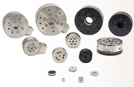
A device that measures the outputting forces and torques from all three Cartesian coordinates (x, y, and z). A six-axis force/torque transducer is also known as a multi-axis force/torque transducer, multi-axis load cell, F/T sensor, or six-axis load cell.
Measure all six components of force and torque in a compact, rugged transducer.
The ATI Multi-Axis Force/Torque Sensor system measures all six components of force and torque. The system consists of a transducer, shielded high-flex cable, and intelligent data acquisition system, Ethernet/DeviceNet interface or F/T controller. Force/Torque sensors are used throughout industry for product testing, robotic assembly, grinding and polishing. In research, our sensors are used in robotic surgery, haptics, rehabilitation, neurology and many others applications.
ATI offers two types of sensing technologies:
• Force/Torque Sensor
• Capacitive F/T
The ATI Multi-Axis Force/Torque Sensor system measures all six components of force and torque. It consists of a transducer, interface electronics and cabling.
Transducer
The compact and rugged monolithic transducer uses silicon strain gages to sense forces. The transducer’s silicon strain gages provide high noise immunity and allow high overload protection, which is standard on all models.
Built-In Capabilities
The Net F/T, DAQ F/T, Controller F/T, and TWE F/T each provide a variety of powerful functions:
• Tool transformations translate and/or rotate the F/T reference frame.
• Demo software allows configuration and basic data logging capabilities.
• Biasing provides a convenient way to offset tool weight.
• Increased system throughput is possible by reducing the number of axes of output. (Not applicable to the Net F/T or TWE F/T.)
• Threshold detection eases integration into industrial applications (Net F/T and Controller F/T only).
• Integral temperature compensation insures accuracy over a wide temperature range.
New Technology – Capacitive F/T
ATI also offers the Capacitive F/T, a Six-Axis Force/Torque Sensor that utilizes capacitive technology in a simple structure, providing a low-cost solution. The Capacitive F/T includes integral overload protection and is IP65-rated for protection against dust and water spray. The sensor is easily connected to computer systems using the optional USB or Ethernet conversion cables. More information here.
The PLUTO-2 phase only Spatial Light Modulator (SLM) consists of a driver unit with standard digital video interface (HDMI) and a phase only LCOS (Liquid Crystal on Silicon) microdisplay with full HD resolution (1920 x 1080 pixel) and 8 µm pixel pitch leading to an active area diagonal of 0.7â with aspect ratio of 16:9.
The PLUTO-2 SLM is a plug & play phase modulator device and can be addressed with phase functions via standard graphics cards as an extended monitor device. The green color channel of the video signal is used for addressing 8 bit gray level patterns (the SLMs native resolution need to be addressed). Addressing can be done using the supplied Pattern Generator software or the SLM Slideshow Player software or standard image viewer software. HOLOEYE also provides an SLM Display Software Development Kit (SDK) which provides APIs (Application Programming Interface) for different programming languages.
HOLOEYE offers different display versions that can be driven with the PLUTO driver unit. The different panel versions are optimized for the use at different wavelength ranges and different applications. All versions use fast full digital addressing which assures high reliability and a compact driver unit as basically only two voltages need to be generated.
The PLUTO-2 driver uses an HDMI interface for addressing phase functions and an USB connection to communicate with the driver (to changing the voltage vs. gray level distribution (gamma control) and dynamic range (voltage across the LC cell) in order to calibrate the SLM for different wavelengths). Besides this the driver features a trigger sync output to synchronize the device with external devices.
The PLUTO-2 driver also features a dual-core ARM® Cortexâ¢-A9 processor which also includes on-chip memory. This enables the user to program additional functionality which is directly processed on the PLUTO-2 device (e.g. a slideshow from images which are loaded from USB flash or from internal memory). The dual-core system runs an embedded Linux⢠SMP operating system and includes a library which provides full control and supervision of the display and driver board. The PLUTO-2 provides access through Serial and Ethernet-over- USB2 (RNDIS) interfaces and can be programmed using standard Ubuntu⢠cross compile GCC toolchain.
The standard PLUTO displays show a reflectivity of approx. 65%-95% (dependent on version) and diffraction efficiencies of more than 80%. Thereby a total light efficiency of more than 50% per addressable diffractive device is possible.
PLUTO Spatial Light Modulator â Microdisplay Features
The LC 2012 Spatial Light Modulator is based on a transmissive Liquid Crystal (LC) microdisplay with 1024 x 768 pixel resolution. The SLM provides a phase shift of about 2 Ï at 450 nm, about 1.8 Ï at 532 nm and around 1 Ï at 800 nm. The microdisplay and drive electronics are packaged into a compact box for easy integration into optical setups. The LC 2012 is addressed using a standard HDMI interface and brigtness and contrast settings can be performed using an USB interface. The LC 2012 Spatial Light Modulator can be used for phase (phase mostly) and amplitude modulation applications dependent on the configuration.
LC 2012 Spatial Light Modulator â Microdisplay Features