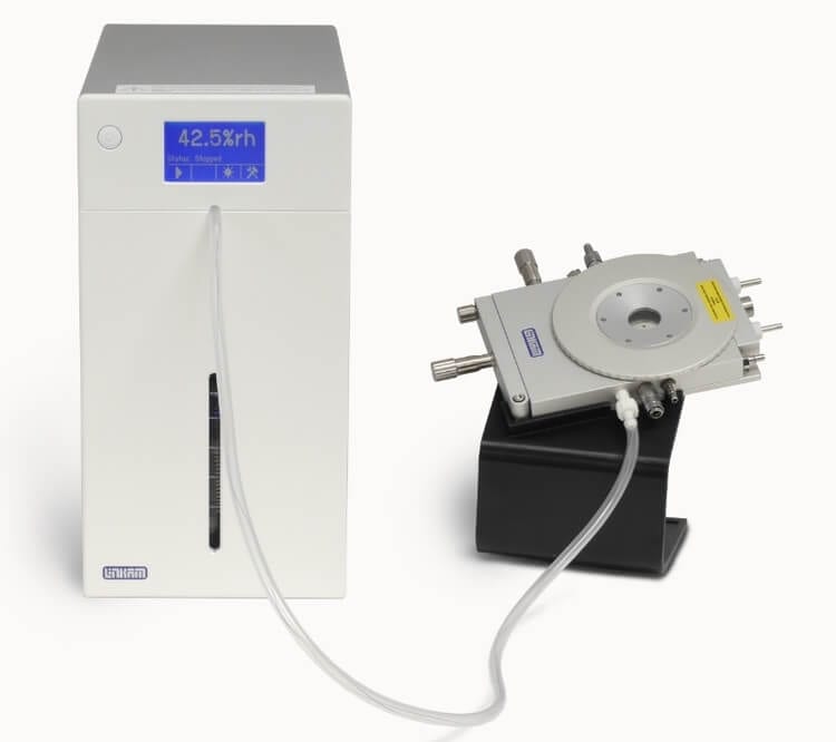
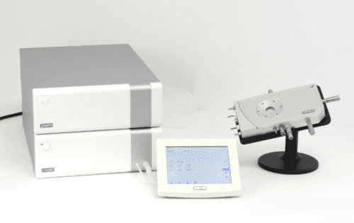
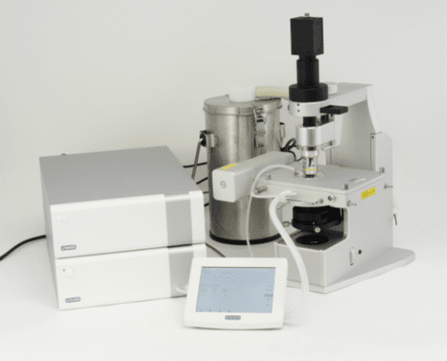
The RH95 quickly controls humidity inside a linkam stage or any other sealed chamber up to a volume of 2000cc from 5 to 90%.
Linkam RH95 Humidity Controller
Unlike other humidity systems, no costly dry air supply is required. Ambient air is dried through a specially designed automatic recycling desiccant system and so can be left controlling humidity for months at a time.
Rather than simply monitoring the humidity of the air supplied to the chamber we also place a sensor inside the chamber to create a feedback loop to the controller.
The LTS420 hotstages are optimised for isothermal sample analysis applications where high speed heating and cooling are compromised by larger sample area with excellent thermal stability of less than 0.1°C.
Linkam LTS 420
The LTS420 is an easy to use, versatile heating and freezing stage. The stage consists of a large area temperature controlled element with a platinum resistor sensor embedded close to the surface for accurate temperature measurements in the range of -196 to 420°C (when used with LNP95 cooling pump).
The sample is simply mounted on a standard microscope slide in direct contact with the heating element and can be manipulated 15mm in X and Y direction. The sample chamber is gas tight and has gas valves to purge with either inert gas or humidity.
This stage is also available with internal electrical connections and probes.
The Linkam Optical Shearing System (CSS450) allows structural dynamics of complex fluids to be directly observed via standard optical microscope while they are under precisely controlled temperature and various shear modes.
Linkam CSS450 Optical Rhelogy Systems
Using the optical shearing cell, it is possible to study the microstructure evolution of complex fluids in great details for many physical processes, e.g. coarsening of binary fluids during their phase separations, flow-induced mixing and demixing of polymer blends, defects dynamics of liquid crystals, aggregation of red blood cells and their deformation in flows etc. We are then in a good position to correlate micro structural dynamics with rheological data for gaining insight into rheology of complex fluids.
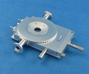
The THMS350V stage enables the user to create low pressure environment during complex heating and cooling profiles. Pressure is measured directly on the sample chamber and displayed in software or on the T95-LinkPad LCD screen.
Linkam THMS350V
It is now possible to carry out ultra low temperature experiments with virtually no gas or air contamination of the sample. It is also possible to first pull a vacuum and then bleed in the desired gas.
A prirani gauge can be supplied to relay the sample chamber pressure to either the LINK software or the LinkPad. By connecting the vacuum pump to the MV196 motorized valve, pressure can be quickly and accurately varied utilizing simple on screen software controls.
To cool samples from ambient down to -196°C, add the LNP95 liquid nitrogen cooling system.
The THMS350V stage enables the user to create low pressure environment during complex heating and cooling profiles. Pressure is measured directly on the sample chamber and displayed in software or on the T95-LinkPad LCD screen.
Linkam THMS350V Ellipsometer
Samples loaded onto a 0.17mm cover slip are placed on a nickel palted heating element to ensure excellent heat transfer and extremely sensitive temperature measurement. A platinum resistor sensor, accurate to 0.01°C provides far more accurate and stable temperature signal that can be achieved with a thermocouple.
Samples can be quickly characterized by heating to within a few degrees of the required temperature at a rate of up to 30°C/min with no overshoot, then slowed down to a few tenths of a degrees per minute to closely examine sample changes. The entire experiment can be saved as an online plot or exported to a spreadsheet application.
The TST350 is built with two precision ground stainless steel lead screws to maintain perfect uniform vertical and horizontal alignment
Tensile Stress Testing System 350
Sample jaws move in opposite directions to maintain sample in both reflected or transmitted microscope fields of view. This also means other transmitted techniques such as x-ray, needed for internal observation of sample structure can be used.
As is expected of Linkam equipment, temperature control and accuracy is second to none, with a range from -196 to 350°C with 0.1°C control and up to 60°C/ min rates, there is virtually no temperature feedback to the measurement of force. The sample chamber is gas sealed and can be controlled with various gases via the gas valves built onto sides of the stage, as well as combined with our RH95Humidity Generator to test samples under different humidity levels.
For general heating and freezing applications in the x-ray spectrometer the HFS based systems have low space requirement , high temperature stability and are ideal for horizontal or vertical mounting.
LINKAM HFSX350 Heating and Freezing System
Two variants are available, the HFSX350-CAP supplied with a 1.7mm capillary tube passing through the heating block for liquid samples, and the HFSX350-GI with flush-mounted heating block for grazing incidence and surface mounted capillary.
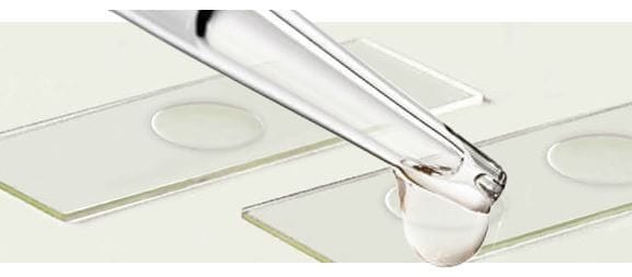
Immersion oils are transparent oils that have specific optical and viscosity characteristics necessary for use in microscopy
Immersion Oil for Microscopy
Immersion oil is used to increase the resolution of a microscope by immersing both the objective lens and the specimen in a transparent oil of high refractive index
Nikon manufactures two types of Immersion Oil for microscopy these being Type A and Type NF. These oils are tested using Nikon objectives and therefore the performance cannot be guaranteed against lenses manufactured by other companies. The refractive indices of other manufactured oils may differ which will impair the results gained from using these with Nikon objectives. It is also important to note that oils should not be mixed as this will impair the performance.
Type A
is general purpose oil used for imaging applications requiring oil immersion, which can incorporate techniques such as brightfield, darkfield, phase and fluorescence. It is available in three sizes of 8ml, 50ml and 500ml, all supplied with a pipette for dispensing.
Type NF
is considered to be the superior grade specifically designed for the ever growing need to improve signal to noise ratios in fluorescence microscopy, particularly in low-light applications or those between 340-380nm, typically associated with calcium or deep UV imaging. The improved quality is due to the type of raw materials used in the oil which relates to a subsequent reduction of auto fluorescence caused by minerals in the oil. There is one size available at 50ml, which is supplied with a plastic pipette for dispensing. It is slightly more viscous and has a slight odour.
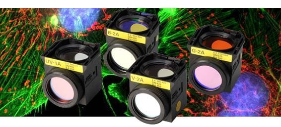
A cube containing the filters and mirror used in epi-fluorescence microscopy to separate fluorescence excitation and emission light
Fluorescent Filter Cubes for Epi-Fluorescence Microscopy
The function of a filter block is to separate fluorescence light returning from the specimen from the light used to excite the specimen so that the fluorescence light can be observed on a dark back ground. The filter block contains an excitation filter, dichroic beamsplitter (mirror) and a barrier / emission filter:
The excitation filter, usually a bandpass filter, allows only wavelengths of light necessary for excitation to pass through to the specimen.
The emission / barrier filter separates fluorescence emanating from the specimen from other background light.
The dichroic mirror separates excitation light from fluorescence by reflecting light of one range of wavelengths and transmitting only light of another range of wavelengths.
The excitation filter, barrier filter and dichroic mirror need to be matched to the excitation and emission characteristics of the fluorescent probe to ensure a high signal to noise ratio between the fluorescence and background light. The ideal combination of barrier filters and excitation filters is one that lets no light pass when combined. However, most filter combinations are not 100% efficient in preventing stray light from reaching the eyes / detector(s). Nikon’s proprietary ‘Noise Terminator’ technology reduces stray light from filter blocks to improve signal-to-noise ratios.
APPLICATIONS
Filter blocks are used in all epi-fluorescence imaging applications. The choice of a specific filter block will depend on the fluorescent probe(s) being used in the imaging application. For a quick and simple-to-use Nikon filter block selector matched to specific fluorescent probes visit:
Introduction to fluorescence microscopy: [microscopyu]
MICROSCOPE CONFIGURATION:
The filter block is housed in a filter cube holder that can be rotated or moved sideways to select the appropriate filter set for imaging. Different microscopes accommodate varying numbers of filter blocks.
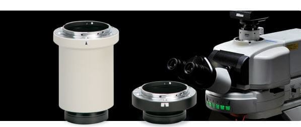
The connection between your microscope and camera
Camera Mounts
There are literally hundreds of potential combinations with many different camera and microscope types. The most fundamental information you will need when deciding which c-mount adapter you need are as follows:
What make and model is your microscope?
This information is important as there will be different focal positions (point at which image data will be collected on the cameras chip) for different manufacturers microscopes. Modern microscopes will tend to have the model type on the microscope body, sometimes near the serial number. As an alternative the microscope manufacturer should be able to identify the microscope by its description or by a photograph.
What make and model is the camera you are trying to attach?
This is an important piece of information as it usually determines the type of thread or fitting the c-mount adapter should have.
What port will you be using?
It is not always possible to connect a camera via a dedicated camera port (sometimes referred to as trinocular tube or beamsplitter). It is important that you identify which port will be used to accept the c-mount.
Is a magnification necessary?
The camera chip (CCD) will be of a certain dimension and will require the appropriate magnification within the c-mount adapter which is sometimes referred to as a relay lens. A good rule of thumb is that whichever size the CCD is as a decimal is what it requires in magnification to fill the viewfield. For example a 2/3” CCD (0.67”) requires a 0.7x c-mount. The CCD size should always be made available by the manufacturer.
It is always remembering that a lower magnification will give a wider field of view and generally brighter images for shorter exposures.