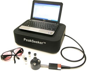
The PeakSeeker Pro represents the state-of-the-art for an accurate, cost-effective and easy-to-use Raman spectrometer. The premier instrument in the Raman Systems line utilizes TE-cooled, high efficiency CCD detector arrays, and is available with either a 532 nm or 785 nm laser.The PeakSeeker by Raman Systems is our most practical and easy-to-use Raman spectrometer. It is an excellent choice for users who want a full featured Raman spectrometer but have a limited budget.
Desktop H-PeakSeeker Pro – Raman Systems Most Versatile System
PeakSeeker Pro is extremely versatile for measuring multiple sample types. The model PRO-785 offers high resolution in the the Raman fingerprint region of organic molecules. See our FAQ answer comparing medium resolution vs. high resolution Raman spectrometers. User friendly software and USB connectivity enables truly plug and play operation. Sampling accessories include a vial holder for liquids and powders. The PRB-HLD is a rack and pinion track stand that provides fine vertical adjustment for precision positioning of the fiber optic probe over solid samples placed on its baseplate. The fiber optic probe accesses remote samples and also interfaces to the microscopy accessories; the RSM Microscope and the MSK Microscopy Kit.
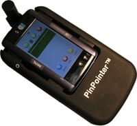
Pinpointer, the “Point-Click” solution designed specifically for quick material identification and verification on the go and for various field deployments.
Portable-PinPointer™ – Newest Handheld “Point, Click, Done” Solution For Material Identification
The PinPointer packs a full-featured Handheld Raman system in a true hand-held unit and is very competitively priced. This is the next generation of the portable Raman spectrometer that was featured in an episode of CSI Miami and a cave exploring documentary by NASA scientists on the National Geographic Channel. The unit is controlled by a miniature Windows computer and features easy-to-use ID-find™ software that provides one-touch identification of unknown substances by immediate comparison of a measured spectrum to the on-board Raman spectral library.

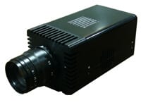
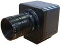
The ARTCAM-407UV-WOM is camera adopting a CCD sensor equipped sensitivity of ultraviolet. The area of visible light is hardly recognized based on its combination with UV lighting equipment and it easily reflects blur, stain and scratch on the surface of objects.
ARTRAY – USB2.0 UV camera (Ultraviolet)
Product Details
High sensitivity in 900nm-1700nm NIR range, storage images with USB2.0 Interface
ARTCAM-031TNIR
*USB2.0 interface, No Capture Board Needed
*Easy to process original images as RAW data are extracted.
*Gain and Shutter Speed are adjustable with enclosed viewer software
*640 x 512 High Resolutions (for ARTCAM-031TNIR)
Product Details
High sensitivity in 900nm-1700nm NIR range, storage images with USB2.0 Interface
ARTCAM-008TNIR
*USB2.0 interface, No Capture Board Needed
*Easy to process original images as RAW data are extracted.
*Gain and Shutter Speed are adjustable with enclosed viewer software
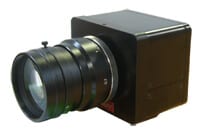
Industrial Camera with USB3.0 Interface, images can be save without compression
ARTCAM-USB3-T2 Series – USB 3.0 interface Industrial Camera
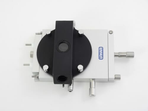
The THMS600 Heating and Freezing Microscope Stage is used in many applications where high heating/freezing rates and 0.01°C accuracy and stability are needed. The THMS600 has a temperature range of -196°C to 600°C..
Linkam THMS600 Heating and Freezing Microscope Stage
Samples can be quickly characterized by heating to within a few degrees of the required temperature at a rate of up to 150°C/min with minimal overshoot, then slowed down to a few tenths of a degrees per minute to closely examine sample changes. The entire experiment can be saved as an online plot or exported to a spreadsheet application.
There are various version options for this stage, including Pressure, vacuum, eletrical sample measurement and sample holders to mount the stage vertically in IRor xray spectrometers.

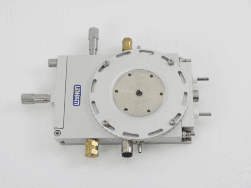
The THMSG600 is based on the design of the highly successful THMS600 stage and then upgraded and modified specifically for geological applications.
The THMSG Geology System is the solution for geologists looking for unrivalled temperature accuracy and control. This precision built hotstage can be found in a many Fluid Inclusion laboratories all over the world.
Unrivalled accuracy and control of temperature enable the user to characterize fluid inclusions to better than 0.1°C and hold a stability of 0.001°C. The THMSG600 has a temperature range of -196°C to 600°C.
The THMS600 is one of the most widely used heating stages on the market, and has now been modified with a special optical adapter to facilitate use on an ellipsometer.
LINKAM THMSEL600 Temperature Controlled Ellipsometer Stage
Samples can be quickly characterized by heating to within a few degrees of the required temperature at a rate of up to 150°C/min with no overshoot, then slowed down to a few tenths of a degrees per minute to closely examine sample changes. The THMSEL600 has a temperature range of -196°C to 600°C.
The THMS600-PS also has a safety release valve that ensures the system cannot be over pressurized.
Linkam THMS600-PS Heating and Cooling Stage
By pressurizing the sample chamber up to 14bar the THMS600-PS stage can be used to investigate the effects of pressure on the sample during heating and cooling experiments. This stage has been used in applications where minimizing sample evaporation and sublimation are required.