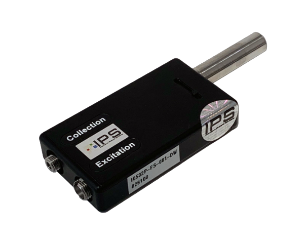
Our Configurable Raman Probe is available in both single or dual wavelength and is optimized to mate with IPS multimode fiber coupled lasers to offer higher throughput and low stray light.
Archives: Product
Andor ZL41 Wave & Cell sCMOS
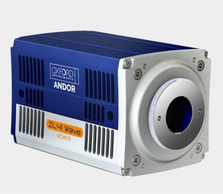
ZL41 Wave and ZL41 Cell is the next generation in the highly successful Zyla sCMOS series.
LuxX+ Diode Laser Series
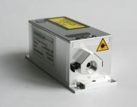
LuxX+® Compact high-performance diode laser modules for biotech and industrial applications
The Omicron LuxX+® Laser Series offers high-perfomance at a compact design. A broad variety of wavelengths and single-mode emission up to 500mW cover a wide range of applications. Easy integration into existing or future designs is assured by versatile input signal types. The USB2.0 and the RS-232 interface support deep integration of the lasers into the application´s process.
Single-Mode Digital U-Type Module with Optical Isolator
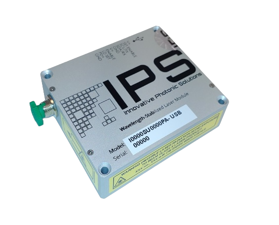
Innovative Photonic Solutions’ proprietary Single-Mode Digital U-Type Module with Optical Isolator has a stabilized peak wavelength that remains “locked” regardless of case temperature (15º to 45º C).
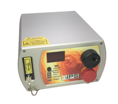
Single-Mode Digital L-Type Module with Optical Isolator
The wavelength stabilized Single-Mode Digital L-Type Module with Optical Isolator can be used in applications such as metrology/interferometry, remote sensing, Raman spectroscopy, direct-diode frequency doubling, and fiber laser seeding.
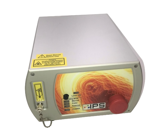
Multi-Mode Digital U-Type Module
Our proprietary multi-spatial-mode wavelength stabilized Multi-Mode Digital U-Type Module features high output power with ultra-narrow spectral bandwidth.
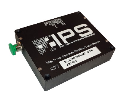
Multi-Mode Digital M-Type Module
Our proprietary Wavelength Stabilized Multi-Mode Digital M-Type Module features high output power with narrow spectral bandwidth. The laser’s stabilized peak wavelength remains “locked” regardless of case temperature (10 to 35 deg. C). Devices can be spectrally tailored to suit application needs and offer side mode suppression ratios (SMSRs) better than 40 dB, thereby providing extremely high signal to noise ratio and making these sources ideal for Raman spectroscopy and pump laser applications.
Tomocube HT-X1 Holotomography
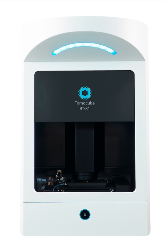
By capturing the intrinsic refractive index (RI) of cells using a low level of light intensity, Holotomography has emerged as a unique solution for live cell imaging that surpasses the compromise between obtaining high image quality and maintaining healthy cells.
Observations of cellular morphology and activity are typically based on labeling of target molecules. These methods are invasive, affect the nature of the target molecules and potentially interfere with their biological relevance. In addition, these forms of label are not effective in providing quantitative information. The tools used to visualize labels are generally damaging to cells as often laser-based illumination is used to excite fluorochromes which in turn causes phototoxic damage to the cells.
The “holy grail” of imaging is to visualize cells without labeling to ensure the cells behave and grow normally. Holotomography (HT) does this by capturing the intrinsic light scattering properties of cellular materials using very low levels of light intensity, just enough to allow the light to pass through the cell. In doing so, the refractive index (RI) information of structures within the cell can be collected and selectively pseudo-colored to reveal the cell and its organelles.
In capturing the RI distribution, the Tomocube HT-X1 Holotomography can also provide quantitative data in 3D such as the volume, surface area, and dry mass of cells and their intracellular structures.
Correlative HT to FL
Holotomography and Fluorescence |
Fluorescence |
Nanoscribe- Quantum X shape
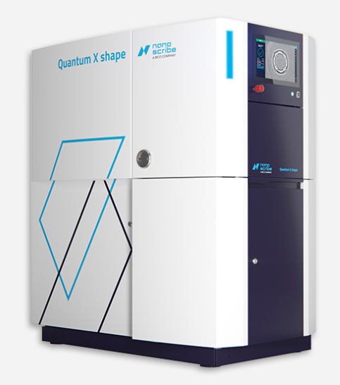
Nanoscribe- Quantum X shape – Tomocube HT-X1 Holotomography – Quantum X shape is a truly capable multi-talent. Based on Two-Photon Polymerization (2PP).
Fastest and most accurate 3D printer for high-end microfabrication tasks
Quantum X shape is a truly capable multi-talent. Based on Two-Photon Polymerization (2PP), the 3D laser lithography system combines proprietary 3D printing technologies that make it the optimal tool for rapid prototyping and wafer-scale batch processing of virtually any 2.5D and 3D shape with submicron precision and accuracy.
Reshaping precision.
As the second high-resolution two-photon lithography system in the industry-proven Quantum X platform, Quantum X shape offers high-resolution 3D Microfabrication capabilities with unmatched precision, next to Nanoscribe’s breakthrough technology of Two-Photon Grayscale Lithography (2GL ®) for surface patterning. The new Quantum X shape’s superior precision relies on the highest voxel modulation rate in class, and an extremely fine address grid, allowing for sub-voxel size shape control. In addition, you benefit from the 2GL voxel tuning capability for 2.5D structures with stunningly smooth, accurately shaped, or micropatterned surfaces.
Reshaping output.
Quantum X shape is the ideal additive manufacturing tool for rapid prototyping of application designs in biomedical devices, microoptics, microelectromechanical systems (MEMS), microfluidics, surface engineering and many more. Wafer handling capabilities make wafer-scale batch processing and small series production of 3D microparts easier than ever.
Reshaping usability.
Control your print job via the device’s integrated touchscreen. Keep an eye on your two-photon lithography system even from the office and in multi-user configurations via nanoConnectX. And benefit from industrial standards and time-efficient wafer batch production.
Technical features in brief
- Rapid prototyping with highest precision and design freedom along a straightforward workflow
- Industry-proven platform for wafer-scale batch processing
- 200 typical mesoscale structures printable overnight
- Print volumes up to 30 cubic centimeters in one pass with the new XLF Print Set
- Broad range of application-specific and universal printing materials
- Open direct laser writing system for custom-made and third-party materials
Nanoscribe – Quantum X align
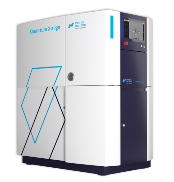
Highest resolution 3D printer with A2PL® technology for nanoprecise alignment
The Aligned 2-Photon Lithography (A2PL®) system Quantum X align enhances Nanoscribe’s field-proven 3D Microfabrication technology based on Two-Photon Polymerization (2PP) by adding high-precision alignment capabilities for highly accurate placement of printed structures. With A2PL freeform microoptics can be printed precisely aligned to the optical axes of fibers or photonic chips with submicron accuracy using this highest resolution 3D printer with nanoprecision aligned 3D printing capabilities. Produce efficient optical interconnects for photonic integration and photonics packaging or miniaturized imaging optics, e.g. for minimally invasive endoscopy.
Technical features in brief
- High-performance 3D Microfabrication by Aligned 2-Photon Lithography (A2PL)
- 3D printing on fibers: Precisely aligned printing on the facets of optical fibers based on fiber core detection
- 3D printing on chips: Precisely aligned printing on the surface or facet of chips based on 3D substrate topography mapping
- 3D alignment: Automatic detection and compensation of substrate tilt in 3 rotation axes
- Smart slicing for high-speed microfabrication