Enquiry Form
Enquiry Form

Upright Microscope Eclipse Si

Focus in comfort. Comfort in focus.

Nikon has designed the ECLIPSE Si to meet the rigorous demands of professionals who spend hours using the microscope. The ECLIPSE Si is ergonomically designed to enhance operational efficiency. This powerful instrument helps you to stay focused for longer by reducing the strain on your body. The ECLIPSE Si is the new standard in microscopes, expanding the possibilities of exploring the micro world.


Upright Microscope Eclipse Ei

Look into the future

Curiosity – the genuine desire to study the world and unlock its mysteries – is the starting point of science and technology.

The ECLIPSE Ei educational microscope offers unique digital and design solutions to ensure the smooth progress of your courses. Using the ECLIPSE Ei to foster your students’ curiosity and maintain their enthusiasm for learning, you can unlock their potential and open their eyes to the world around them.

Nikon Smart Imaging System Eclipse Ji

Research Microscope Power in a Benchtop Assay Instrument

Simple Operation

Minimal experimental setup and complexity by AI-driven assays and analysis.

Easy Cellular Imaging

No need to master complicated microscope hardware and software – ECLIPSE Ji makes data collection and cellular imaging assays easy.

High Quality Optics

Renowned Nikon optics provide clear and sharp images on plate assay devices.

Inverted Research Microscope Eclipse Ts2R

A compact inverted research microscope configurable with a wide variety of observation methods

A new intermediate-sized inverted research microscope designed for a wide variety of applications. Do more than before!
Using these innovative optics and a fly-eye lens, crystal clear fluorescent images with improved S/N ratios are possible, even in low-excitation light applications such as cell division. Additionally, even at low magnifications, the SMZ25 microscope has a 35mm F.O.V, allowing users to capture an entire 35mm dish with uniform brightness.

The Eclipse Ts2R is a compact inverted research microscope that is configurable with a wide variety of observation methods. The broad range of observation methods, smaller body and improved ease of use will directly benefit research laboratories and improve workflow.

In addition to conventional contrast methods, the Eclipse Ts2R features versatile observation methods, including Phase Contrast, DIC, Hoffman Modulation Contrast and Emboss Contrast. It is also compatible with a selection of high performance and quality optical accessories to function similarly to the inverted research microscope Eclipse Ti and produce clear, sharp images.

Inverted Routine Microscope Eclipse Ts2

Nikon’s all-new inverted routine microscope featuring a compact footprint, newly developed contrast observation method and improved operability.

New compact inverted routine microscope that does more than ever before!
Utilizing Nikon’s industry-leading CFI60 optical system and LED illumination for both diascopic and epi-fluorescence imaging, the new Eclipse TS2 microscope offers brilliantly clear images, enabling more efficient cell culture observation and documentation. Furthermore, a newly developed contrast observation method and streamlined operation enhance its use for routine laboratory work.

Microscope System for ICSI – Eclipse Ti2-U IVF

Superior optical performance and precision for ICSI

Nikon microscopes have played a significant role from the early days of Intracytoplasmic Sperm Injection (ICSI) and continue to contribute to the advancement of this field through superior optical performance and precision.

The ECLIPSE Ti2-U IVF provides an ultra-stable platform for performing precision manipulation procedures such as ICSI. The intermediate magnification switching function (1.5X) allows easy switching of observation magnifications without changing objectives.

Super-Resolution Microscope N-STORM 4.0

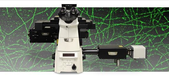
All-new super-resolution microscope featuring acquisition speeds ten times faster than conventional N-STORM systems.

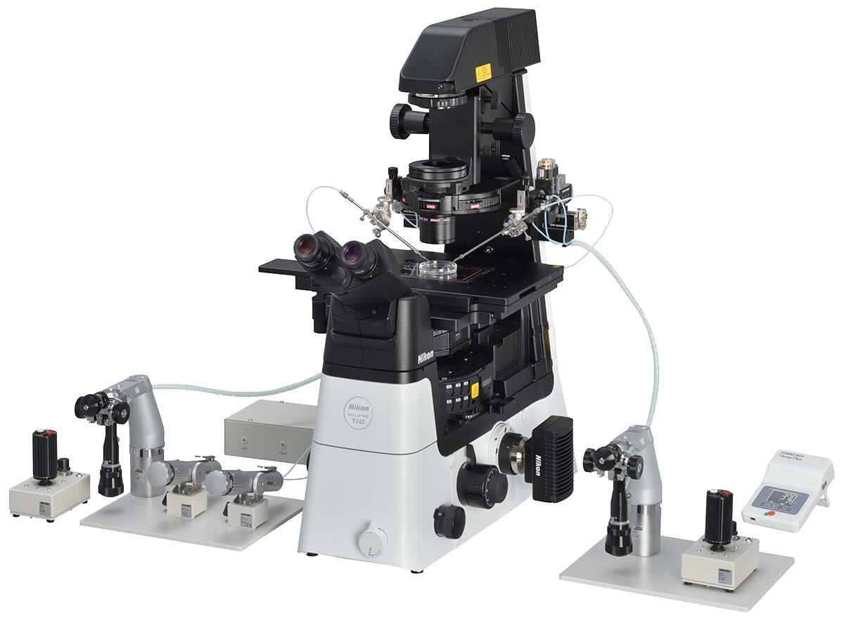
Nikon’s Next Generation N-STORM Super-Resolution Microscope System.
With ten times faster acquisition speeds compared to conventional N-STORM, N-STORM 4.0 enables imaging of live cells with nanoscale resolution.

Nikon has established a leading position in the field of super resolution microscopy since the release of N-STORM in 2010. STORM, or Stochastic Optical Reconstruction Microscopy is a super-resolution technique developed by Dr. Xiaowei Zhuang, a Howard Hughes Investigator at Harvard University. The technology localizes temporally isolated fluorescent molecules to reconstruct images with nanoscale resolution (~10 times greater resolution compared to conventional light microscopy). N-STORM 4.0 is the next step in the evolution of STORM imaging, enabling STORM imaging of dynamic processes in live cells.

N-STORM 4.0 utilizes an improved laser excitation design and a sCMOS camera that dramatically improves acquisition rates.

There is a rapidly growing interest in super-resolution microscopy as evidenced by the recent 2014 Nobel Prize in Chemistry awarded to the field of super-resolution microscopy. There is also an increasing need to expand these techniques to broader applications like live-cell imaging. Recent studies show that localization-based super-resolution imaging of live cells can provide unprecedented insights into the nanoscale dynamics of intracellular structures such as focal adhesions1, transferrin clusters in clathrin-coated pits2, and organelles including mitochondria and endoplasmic reticulum3.

N-STORM 4.0 utilizes an improved laser excitation design and a sCMOS camera to improve acquisition rates from minutes to seconds while gathering incredibly high molecule counts. In addition, the new system offers expanded flexibility in the imaging field of view, allowing users to choose the best suited mode for their application, whether it be high-speed imaging of live cells or gathering large fields of view of fixed cells.

N-STORMv4-camera
N-STORM 4.0 utilizes an improved laser excitation design and a sCMOS camera that dramatically improves acquisition rates.

1 Shroff, H. et al., 2008, Nature Methods, 5 (5), p417-423.
2 Jones, S.A. et al., 2011, Nature Methods, 8 (6), p499-505.
3 Shim, S-H. et al., 2012, Proc. Natl. Acad. Sci., 109, p13978-13983

Super-Resolution Microscope N-STORM

Tenfold increased resolution in x, y and z.

STochastic Optical Reconstruction Microscopy (STORM) reconstructs a super-resolution fluorescence image by combining precise localization information for individual fluorophores in complex fluorescent specimens. N-STORM takes advantage of Nikon’s powerful Ti2-E inverted microscope and applies high-accuracy, multi-color localization and reconstruction in three dimensions (xyz) to enable super-resolution imaging at tenfold the resolution of conventional light microscopes (up to approximately 20 nm in xy).

This powerful technology enables the visualization of molecular interactions at the nanoscopic level, opening up new worlds of scientific understanding.

Super-Resolution Microscope N-SIM E

All-new personal structured illumination super-resolution system for the individual lab.

Introducing an all-new super-resolution system for the individual lab. Achieve 2x resolution at half the price!
Utilizing structured illumination microscopy (SIM) technology, the all-new N-SIM E realizes double the spatial resolution of conventional optical microscopes (to approximately 115 nm). N-SIM E is a streamlined, affordable super-resolution system supporting only essential, commonly used excitation wavelengths and imaging modes, making it an obvious choice for individual labs.

PicoQuant Time-resovled Confocal Fluorescence Microscope – MT200

Time-resolved Confocal Fluorescence Microscope with Unique Single Molecule Sensitivity

Picoquant Time-resolved Confocal Fluorescence Microscope – MicroTime 200

  • Complete confocal system with laser combining unit, inverted microscope body and multichannel detection unit
  • Turn-key diode lasers for multicolor excitation from 375 to 900 nm
  • Up to 4 truly parallel detection channels using application-optimized detection with SPADs, PMTs or Hybrid-PMTs
  • Time-Correlated-Single Photon Counting (TCSPC) and TTTR mode for investigation of fast dynamics in FCS and FLIM
  • Piezo scanning for 2D- and 3D-lifetime imaging and accurate point positioning
  • Two optional exit ports for additional hardware, e.g. spectrographs
  • Advanced easy-to-use data acquisition, analysis and visualisation software SymPhoTime 64
  • Unique upgrades to 2focus FCS, simultaneous AFM/FLIM, UV measurements
  • STED add-on for super-resolution imaging